


Sciatica and progressive numbness: why not wait?
17/08/2026
Sciatica and calf pain after driving
17/08/2026
IN BRIEF
| In brief, disc herniation and heel pain related to the S1 root describe a radiculopathy causing pain that radiates down to the heel and the outside edge of the foot. In Montreal, this service helps connect symptoms, examination, and, if useful, imaging to guide appropriate management and the maintenance of activities. Key benefits include a better understanding of the pain pathway and motor/sensory signs, allowing for effective non-surgical approaches and monitoring of progression. The approach relies on a concordance between the pathway, strength, and reflexes; imaging is only used if it can change the therapeutic decision. In cases of new weakness, rapid deterioration, or signs of urgency, a quick consultation is recommended. |
Brief summary: Disc herniation and heel pain – possible involvement of the S1 root.
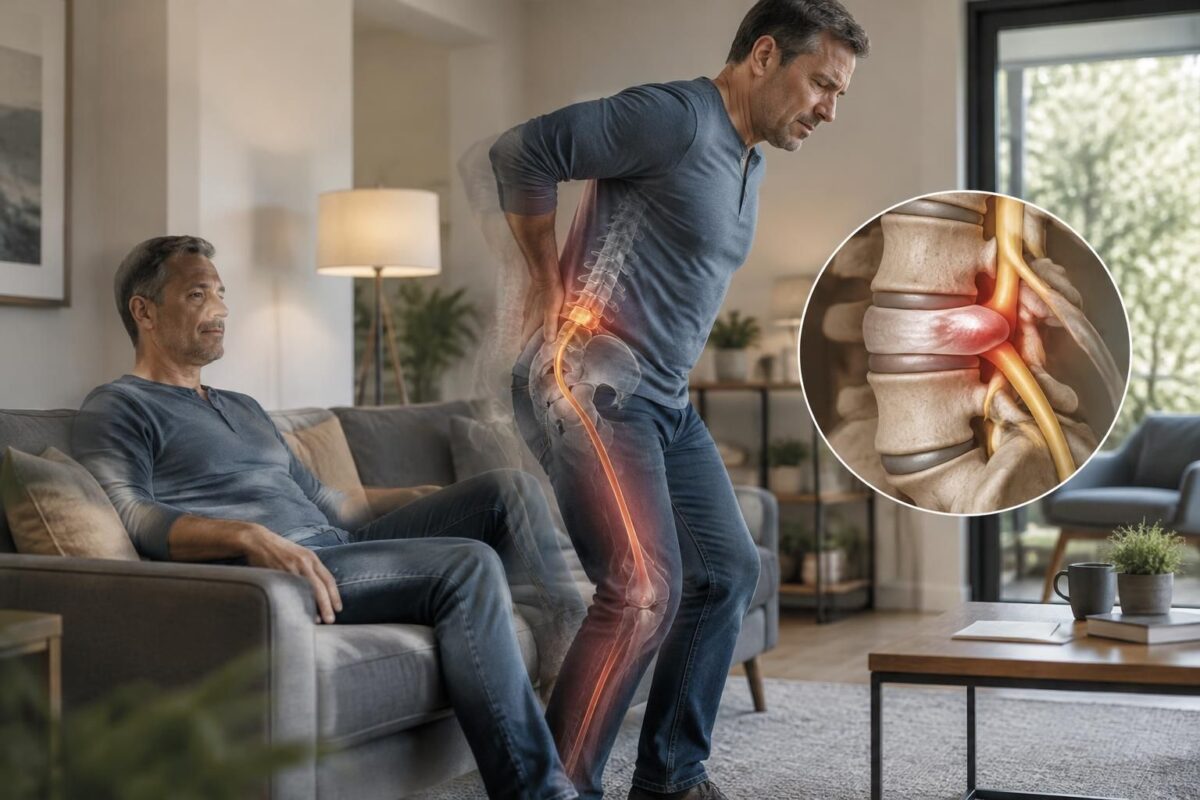
- A L5–S1 disc herniation can irritate the S1 root, causing pain that follows the posterior pathway of the leg down to the heel and the outside edge of the foot, sometimes extending to the calf.
- Typical signs include neuralgic pain along the S1 pathway, weakness in plantar flexion, and difficulties in tiptoeing, with a potentially diminished Achilles reflex.
- The diagnosis relies on pain-pathway concordance, function, and neurological examination, and imaging is interpreted in conjunction with the clinical picture.
- The presence of a herniation on MRI alone does not explain the symptoms; other causes may exist and treatment is often focused on inflammation and non-surgical measures.
A quick assessment can help confirm the concordance between signs and imaging and suggest the best management options.
Quick summary: This article explains how a disc herniation can affect the S1 root and cause heel pain. We describe the nerve pathway, clinical signs, assessment, and non-surgical management options, with practical guidelines to inform medical decision-making.
Heel pain may arise from an issue with the S1 root, which contributes to the sciatic nerve. The pain pathway can descend behind the thigh and calf to the heel, to the sole or the outside edge of the foot. Radicular irritation may be accompanied by weakness in plantar flexion and an extended time needed to walk on tiptoes.
The diagnosis relies on the concordance between symptoms, function, neurological examination, and, when relevant, imaging. A disc herniation visible on MRI is only relevant if the level, side, and clinical picture agree. Progressive weakness, a drop foot, or signs of cauda equina syndrome change the evaluation priority.
Clinical markers around the S1 root
- Marker 1: The S1 root is often associated with the back of the calf, the heel, and the outside edge of the foot.
- Marker 2: Weakness in plantar flexion can make it more difficult to stand on tiptoes on the affected side.
- Marker 3: The Achilles reflex may be decreased, but no isolated sign confirms an S1 radiculopathy.
The pain pathway is evaluated by comparison with motor, sensory, and functional signs. A hernia cannot alone be considered the cause of symptoms. Disc abnormalities may exist in individuals without pain.
Essential elements of the evaluation
- Pathway: precise location of pain, paresthesias, and areas of numbness.
- Strength and function: comparison of key movements and gait.
- Imaging: linking the level and side of the discopathy with the clinical picture, rather than isolated examination.
- Progression: monitoring of symptoms and defining criteria that might otherwise guide management.
For more information on the link between sciatica and limb pain, you can refer to the following resources:
Sciatica and numbness in the leg and
L5-S1 disc herniation and sciatica.
Imaging and management
Imaging must be interpreted in relation to the clinical picture. A level, a side, and concordant clinical signs reinforce the diagnosis. Targeted imaging is useful when the result can modify management, rather than as a routine examination.
Neurovertebral decompression schemes and other non-surgical approaches exist, but these must be presented transparently regarding the uncertainty of data and not promoted as guaranteed standard treatments. For more information on non-surgical options and their framework, refer to specialized resources available on the following reference sites.

Disc bulge and pain after exertion
IN BRIEF A disc bulge is a disc that slightly protrudes between two vertebrae; after exertion, it may be associated with low back pain, sometimes radiating into the leg. An imaging result alone is not enough to explain the pain…
Pain trajectory and treatment options
Pain associated with S1 involvement can be described as a burning sensation or electric shock that follows the nerve pathway. The treatment favors a multimodal approach tailored to each patient, aiming to improve function sustainably rather than aiming for a temporary reduction in pain.
Additional information and clinical perspectives on sciatica and the nerve pathway can be found in the following resources:
Tingling and nerve trajectory and
Extruded disc herniation and clinical significance.

Disc bulge and tingling in the foot
IN SHORT A disc bulge refers to a disc that bulges beyond its boundary; if it irritates a nerve root, it can contribute to tingling in the foot, numbness, or pain in the leg. However, MRI alone is not sufficient…
When to consult quickly or in an emergency?
Consult quickly if the following occur:
- New, significant, or rapidly progressive weakness.
- Recent foot drop or deteriorating gait.
- New urinary or bowel disturbances.
- Numbness in the saddle area or perineum.
- Significant bilateral symptoms, fever, major trauma, or general deterioration.
In these cases, do not wait for a routine appointment.

Disc bulge and intermittent numbness
IN SHORT A bulging disc associated with intermittent numbness may sometimes indicate nerve irritation, but an MRI alone does not allow determining the cause. Connecting the symptoms’ path, sensitivity, and strength helps guide the evaluation and next steps. New or…
Stay active and management trajectory
Clinical recommendations advocate for self-management and maintaining normal activities as much as possible, with adaptations according to the needs and abilities of each individual. Imaging and clinical decisions should be guided by the concordance between the painful trajectory and neurological and functional signs, rather than by the mere presence of a herniation on the MRI.
For additional information on non-surgical approaches and indications for decompression, see the following resources:
- Sciatica and mild lower back pain but very painful leg
- L5-S1 disc herniation and sciatica
- Sciatica and foot pain in the car
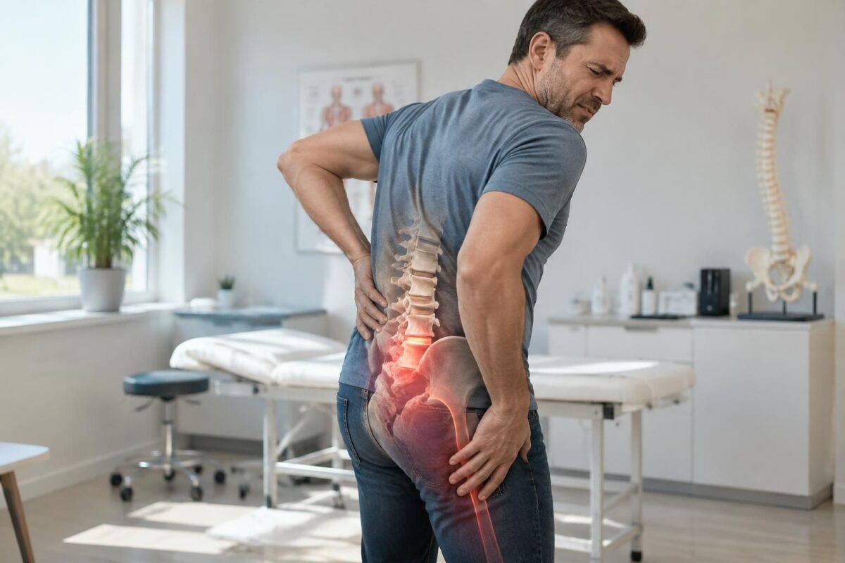
IN BRIEF A disc bulge is a diffuse protrusion of the disc and does not always cause symptoms. A buttock pain may sometimes be associated with nerve irritation, but the bulge visible on imaging is not enough to establish the…
Resources and Useful Links
For additional information and clinical perspectives, see:
- L5-S1 disc herniation and sciatica (TAGMED Clinic)
- Tingling and nerve pathways in Montreal
- Extruded disc herniation and clinical significance
- Sciatica and numbness in the leg
- Disc herniation – symptoms and treatment
- Carpal tunnel syndrome
- Neurovertebral decompression
Limited professional and training reference: NUMSS, NAO, National University of Medical Sciences, National Academy of Osteopathy.
Medical disclaimer: The information and advice provided on this site are not a substitute for the opinion, diagnosis, or treatment of a healthcare professional. Please note that Sylvain Desforges, B.Sc., D.O., N.D., Doctor of Osteopathy, is neither a medical doctor nor a physician and is not a specialist in a medical specialty as defined by the Collège des médecins du Québec. Manual medicine, functional medicine, and sports medicine as described on this site exclude any medical treatment or diagnosis made by a physician or specialist. Always consult your doctor for any medical questions. For more details, please read our full Legal Notice.
- L5-S1 disc herniation may irritate S1 root and generate pain that descends to the heel, the sole, or the outer edge of the foot.
- The painful trajectory typically follows the S1 territory with pain behind the calf and the heel.
- Examination: Achilles reflex may be diminished; plantar flexion weakness may hinder rising on toes.
- Note: a hernia may be present without necessarily accounting for the pain; imaging must be interpreted with the clinical picture.
- Evaluation: look for concordance between trajectory, strength, sensation, reflexes, and function.
- Imaging: useful only if it can modify management; relate the level and the side to the picture.
- Emergencies: new/progressive weakness; foot drop; urinary or bowel disturbances; saddle numbness; fever or major trauma.
- Management: stay active, self-management, and activity adaptation; prioritize non-surgical approaches and inform about uncertainties.

This recommendation explains how a L5-S1 disc herniation can affect the S1 root and cause pain that descends to the heel. It details the painful trajectory, associated neurological signs, the importance of imaging in context, and non-surgical management options, with clear guidelines to guide evaluation and treatment decisions. It emphasizes the concordance between symptoms, examination, and imaging rather than an isolated interpretation of a scan.
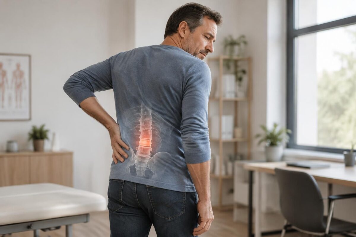
Disc herniation and lumbar stiffness
IN BRIEF The disc bulge is a protrusion of the disc between the vertebrae, without a necessary tear, and does not always indicate a herniation or a cause of pain. It can accompany lumbar stiffness, but the link must be…
Understanding pain related to S1 and the painful trajectory
An L5-S1 disc herniation can irritate the S1 root and generate pain that descends along the back of the leg to the heel, to the outer edge of the foot, or to the sole. The Achilles reflex and the ability to stand on tiptoe can aid in assessment and estimating neuromuscular status. The painful trajectory is often accompanied by paresthesias or numbness and can be exacerbated by prolonged sitting or during exertion.
The pain radiating to the thigh and calf may be more pronounced depending on which root is affected. The plantar flexion test and walking on tiptoes help to reveal any potential weakness in the affected limb.
Quick Guidelines
Landmark 1 The S1 root is often associated with the back of the calf, the heel, and the outer edge of the foot.
Landmark 2 A weakness in plantar flexion can make rising onto the toes more difficult on the affected side.
Landmark 3 The achilles reflex may be diminished, but no isolated sign allows for the confirmation of S1 radiculopathy.
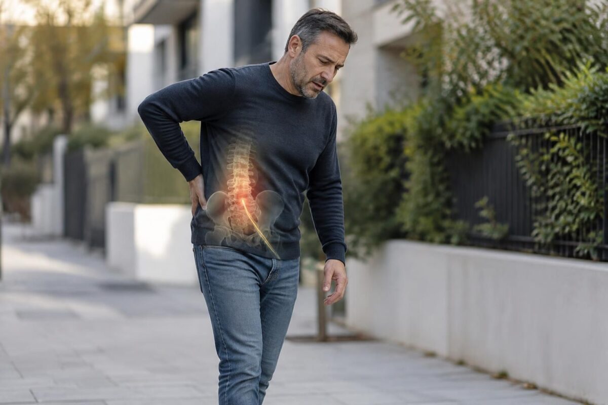
Disc herniation and pain while walking
IN BRIEF A disc bulge is a protrusion of the disc between the vertebrae, which can sometimes be accompanied by pain while walking or radiating down the leg. A gentle and progressive walk helps maintain activity if symptoms remain stable,…
Imaging and clinico-imaging concordance
A disc herniation visible on MRI is only relevant if the level, side, and clinical picture match. The presence of a herniation alone is not enough to explain the symptoms, and disc abnormalities can exist in pain-free individuals. Imaging must be interpreted in relation to the evolution of symptoms and motor and sensory signs.
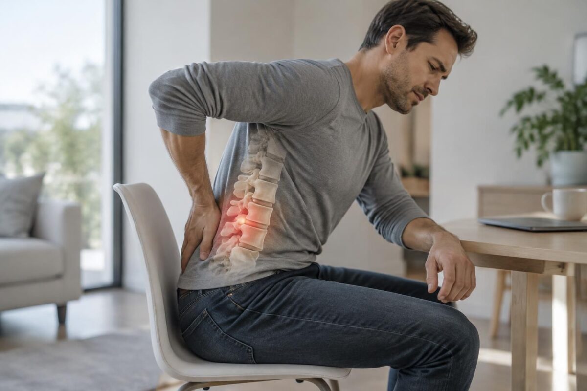
Disc bulge and pain in sitting position
IN BRIEF A disk bulge is a protrusion of a disk between the vertebrae; it does not always cause symptoms. Prolonged sitting can accentuate lower back pain and sometimes pain that radiates into the buttock or leg. Varying positions, moving…
Key assessment elements
Pathway: accurately locate pain, paresthesias, and areas of numbness.
Strength and function: compare key movements, balance while walking, and tasks revealing possible weakness, particularly during plantar flexion and walking on tiptoes.
Imaging: relate the level and side to the entire clinical picture rather than treating an isolated report.
Evolution: track symptoms’ trends and adjust care orientation if necessary.
Disc herniation and fluctuating symptoms
IN BRIEF A disc bulge is a protrusion of the disc between the vertebrae, which may not necessarily cause symptoms. When pain fluctuates, its intensity can vary based on movements, activities, and other factors; the bulge visible on MRI does…
Practical management and recommendations
Staying active is recommended: NICE guidelines advocate for self-management and maintaining normal activities as tolerable as possible, with adaptations according to abilities. Targeted imaging should be used when the result is likely to change management. Lumbar traction is not recommended for low back pain with or without sciatica, and motorized decompression should not be presented as a standard treatment without information on data uncertainty.
Non-surgical management relies on reducing radicular inflammation and strengthening core muscles. Anti-inflammatories and a targeted rehabilitation program can speed up recovery. A proper posture, gradual walking, and specific exercises support stabilization and sustainably reduce pain.
Signs of urgency and when to consult quickly
Consult quickly in case of new or rapid progression of weakness, foot drop, urinary or bowel disturbances, saddle numbness, or significant bilateral symptoms, fever, or major trauma. These signs require prompt evaluation and should not be delayed.
| Element | Brief observations |
|---|---|
| Evaluation axis | Concordance between painful pathway, motor signs, and imaging results for S1 root. |
| Typical pain | Posterior pain in the calf and heel, sometimes the outer edge of the foot. |
| Motor signs | Weakness in plantar flexion and difficulty rising onto tiptoes. |
| Reflexes and sensitivity | Achilles reflex potentially diminished; possible paresthesias. |
| Imaging | MRI must correspond to the clinical level and side. |
| Associated neurological signs | Paresthesias and possible weakness related to S1. |
| Differential diagnosis | Disc herniation can exist without symptoms; clinical correlation necessary. |
| Initial management | Anti-inflammatories and maintaining appropriate activity; avoid prolonged rest. |
| Traction and decompression | Traction not recommended; motorized decompression is not a standard treatment. |
| Emergency and alert signs | Urinary disturbances, rapid weakness, foot drop, or marked bilateral pain require urgent evaluation. |
Testimonials on disc herniation and heel pain that may involve the S1 root
Testimonial 1: Sophie, 41, describes a heel pain that travels up the back of the leg to the calf after a painful episode in the lower back. The picture suggests an L5-S1 disc herniation irritating the S1 root, with paresthesias and sensitivity at the heel and outer edge of the foot. Upon examination, the achilles reflex may have been diminished and plantar flexion limited, which guided the diagnosis. The assessment confirmed the concordance between pain pathway and neurological signs, and imaging clarified the level. Thanks to tailored management combining specific osteopathy and motorized neurovertebral decompression, Sophie regained normal walking and can rise onto tiptoes. If you are experiencing a similar situation, a consultation may help check the concordance between signs and imaging and consider suitable options.
Testimonial 2: Marc, 54, developed lumbosciatica after exertion. The heel pain and radiation towards the calf led to thoughts of an S1 root involvement. Examination showed S1 radiculopathy and imaging confirmed an L5-S1 disc herniation. The achilles reflex was sometimes weak and plantar flexion limited during tests. With a targeted therapeutic plan, followed by adapted exercises, Marc regained his ability to walk on tiptoes and noted a lasting improvement in function. If you find yourself in this situation, a consultation can guide you through the right steps and management options.
Testimonial 3: Claire, 29, suffered from repeated episodes of heel pain that worsened after days on her feet. The pain pathway often corresponded to an L5-S1 disc herniation irritating the S1 root, with numbness at the back of the foot. The assessment revealed strong concordance between the pain location, slight weakness during plantar flexion, and neurological signs. Thanks to tailored management and maintaining compatible activity, Claire resumed her routine without heel pain. If you recognize yourself, considering an evaluation may confirm concordance between symptoms, examination, and imaging, and guide management.
Testimonial 4: Jean, 63 years old, had heel pain associated with discomfort while walking and low back pain. The examination sought to find the correlation between the painful pathway and the motor and sensory signs involving the S1 root. Imaging revealed a disc herniation with dynamic lumbar adjustments. After an approach combining specific osteopathy and decompression techniques, improvement in function became sustainable, and walking returned to his usual level. If this resonates with your experience, a consultation may clarify the next steps.
Testimonial 5: Laura, 45 years old, noted that a painful episode in her heel often occurred with pain radiating up her leg. The diagnosis was an L5-S1 disc herniation involving the S1 root, explaining the heel pain and paresthesias. The assessment linked the painful pathway to the neurological picture, and imaging helped confirm the level. After a personalized program and appropriate rehabilitation, Laura has regained sustainable improvement in her mobility and can resume her daily activities. If you relate, a consultation can clarify the signs to watch for and the management options.
Sylvain Desforges, osteopath and expert in integrative manual medicine, is particularly interested in the mechanisms linking lumbar disc herniation to heel pain via the S1 root. His approach rests on a comprehensive assessment that connects symptoms to function and imaging when pertinent, to establish a personalized and realistic management plan. In his practice, he prioritizes non-surgical options based on available data and, when indicated, the integration of advanced technologies to optimize recovery and patient well-being.
An L5-S1 disc herniation can irritate the S1 root, causing pain that radiates up the back of the leg to the heel, the sole, or the outer edge of the foot. This pain may be accompanied by weakness in plantar flexion and diminished Achilles reflex. The examination focuses on the precise location of the pain, the level of potential weakness, and the muscle control of the muscles responsible for walking and foot stability. The clinical picture may be complemented by simple tests that assess the functionality of the myotendinous chain involved, but no isolated measure is sufficient to confirm S1 radiculopathy.
The effective diagnosis relies on a concordance between the painful pathway, sensory extent, muscle strength, and imaging elements when useful. The physician seeks to link the level and side of the disc damage to the clinical presentation. It is important to remember that the presence of a disc herniation does not alone prove it is the cause of symptoms; disc anomalies can sometimes exist without pain, and the relationship between imaging and symptoms must be established with caution.
In the case of S1-related heel pain, certain markers are useful for guiding the assessment. The S1 root contributes notably to the sensation of the posterior-external aspect of the foot and to the function of the muscles that allow toe pointing. Progressive weakness in plantar flexion may make it more difficult to stand on tiptoe on the affected side, and the Achilles reflex may be diminished, but no single sign is sufficient to confirm S1 radiculopathy. Calf pain associated with paresthesias or reduced sensitivity along the sciatic nerve pathway supports suspicion, but the diagnosis relies on the entirety of clinical data.
The therapeutic approach follows current recommendations that favor self-management and the maintenance of normal activities when possible, while adapting effort and posture. In practice, Dr. Desforges proposes a multimodal approach that may include targeted osteopathic techniques, active maintenance programs, and, when relevant, complementary therapies such as motorized decompression, laser treatments, and shockwaves, always within the framework of transparent information about data uncertainties and realistic goals.
For accurate diagnoses and treatment decisions, it is essential to distinguish radicular causes from local heel pathologies, such as plantar fasciitis or other foot pains. S1 radicular pain is characterized by signs that evolve over time and align with the pathway of the nerve as well as the corresponding motor and sensory domains, rather than by a single localized pain. The assessment seeks to establish a concordance between the painful pathway, strength, sensitivity, reflexes, and imaging results to guide appropriate and effective management.
If you experience heel pain that may be related to S1 radiculopathy, a consultation can help clarify the concordance between your symptoms, your examination, and any imaging, the signs to watch for, and the management options suited to your situation.
Quick summary: an L5-S1 disc herniation can irritate the S1 root, leading to pain that descends the back of the leg to the heel and the outer edge of the foot, with possible weak plantar flexion and diminished Achilles reflex. The assessment relies on the concordance between the painful pathway, motor and sensory signs, and imaging when necessary.
An heel pain associated with sciatica may reflect irritation of the S1 root due to an L5-S1 disc herniation. The pain may descend behind the thigh to the heel, sole, and outer edge of the foot, and may be accompanied by weakness in plantar flexion or difficulty lifting the heel while walking. To better understand this connection, it is useful to examine the painful pathway, the motor signs, and imaging when necessary, while consulting specialized resources such as those dedicated to the association between disc herniation and sciatica. To delve deeper into the mechanisms and options, you may consult additional information sources on informative link on L5-S1 disc herniation and on the relationships between pain and disc herniation.
The S1 localization is frequently associated with the back of the calf, the heel, and the outer edge of the foot. Weakness in plantar flexion may make it more difficult to stand on tiptoe on the affected side, and the Achilles reflex may be diminished. However, no isolated sign is sufficient to confirm S1 radiculopathy; it is the entirety of the clinical picture and its evolution that guide the diagnosis and treatment. To enhance your understanding, specialized resources explain why the presence of a disc herniation alone is not enough to establish unique causality and how inflammation may play a major role in pain.
In the assessment, we look for a match between the pain pathway, muscle strength, sensitivity, and function. This is accompanied by an evaluation of the level and side of the hernia on imaging, if it may alter the management. Resources like those dedicated to assessing the sequestered disc herniation and radiculopathy provide useful benchmarks on the importance of a comprehensive assessment rather than just the radiological report.
The therapeutic approach largely relies on maintaining appropriate physical activity and non-surgical approaches when the initial consultation indicates so. NICE recommendations encourage self-management and maintaining normal activities as much as possible, with adjustments based on abilities and needs. Complementary resources discuss the limits of traction and the relative usefulness of motorized decompression in the context of low back pain with sciatica, emphasizing informed patient information.
In case of deterioration or alarming signs (new or aggravated weakness, walking disturbances, significant bilateral pain, urinary or sensory issues in the perineum), urgent consultation should be considered rather than a routine appointment to avoid potential complications and to quickly direct management.
A lasting improvement in function and the ability to walk is generally more useful than a one-time reduction in pain. Heel pain associated with S1 radiculopathy can evolve favorably with a multimodal approach focused on education, appropriate activity, and non-surgical treatments when appropriate. To delve deeper into the mechanisms and associated therapeutic options, consult specialized resources like Sciatica and lumbar disc herniation – understanding the link and discussions around non-invasive treatments and natural approaches.
To enrich your understanding of the different scenarios surrounding disc herniation and heel pain, you can also consult dedicated insights on L5-S1 disc herniation, including pages explaining symptoms and potential treatments, like L5-S1 disc herniation: symptoms and treatments to consider and resources that examine the links between heel pain and sciatica.
Please note that pain and signs vary among individuals and that radiological results do not always predict pain. For a comprehensive assessment tailored to your situation, a consultation with a healthcare professional is recommended. Other useful resources address specific aspects, such as evaluating painful areas, inflammatory mechanisms, and non-surgical treatment options, available particularly in specialized guides and reviews.

Sylvain Desforges, B.Sc., D.O., N.D., docteur en ostéopathie
Dr Sylvain Desforges, B.Sc., D.O., N.D., osteopath, is the founding president of TAGMED and of the ACMA association. He dedicates his practice to chronic spinal pain, low back pain and symptoms compatible with sciatica, particularly when pain radiates toward the buttock, leg, calf, foot or toes.
Through SOS Sciatique, he publishes educational content on sciatica symptoms, possible causes such as disc herniation, disc narrowing, foraminal stenosis or irritation of a lumbar nerve root, as well as non-surgical approaches that may be considered according to the evaluation. At Clinique TAGMED, care may include, depending on the case, motorized neurovertebral decompression, specific osteopathic care or precision percussion.
For an evaluation related to sciatica treatment in Montreal / Mount Royal or sciatica treatment in Terrebonne, please consult the services available at Clinique TAGMED.
Medical disclaimer
The information published on SOS Sciatique is provided for educational purposes only and does not replace a consultation, diagnosis, opinion or treatment provided by a physician or another authorized healthcare professional. The author of this article is neither a medical doctor nor a specialist in a medical specialty recognized by the Collège des médecins du Québec. The osteopathic, manual medicine or functional support approaches described on this site do not constitute medical acts and are not intended to replace medical follow-up.
Consult a physician or emergency service in cases of loss of bladder or bowel control, anesthesia in the saddle area, significant or progressive weakness in the leg or foot, fever, recent trauma, severe pain or rapid worsening of symptoms. For more details, please read our Legal Notice.