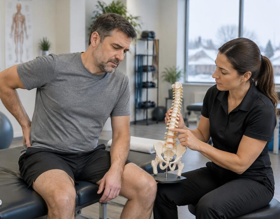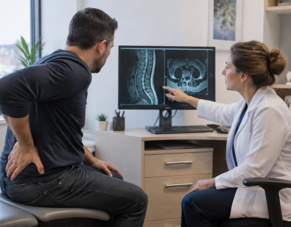



Sciatica in Montreal: understanding the lumbar levels on an MRI
15/10/2026
Sciatica and stomach pain
15/10/2026
IN BRIEF Sciatica may be related to irritation of a lumbar nerve root. A lumbar MRI shows the disks and nerves at different levels, such as L4-L5 or L5-S1, to help identify an abnormality and compare it with symptoms. In Terrebonne, the interpretation of images must be performed by a healthcare professional: an abnormality on the MRI does not, by itself, confirm the cause of the pain. |
A lumbar MRI shows the disks, joints, and nerves of the lower back. The levels are designated from L1-L2 to L5-S1. For a sciatica-type pain, the L4-L5 and L5-S1 levels are often examined, as nerve irritation at these sites can contribute to pain that radiates down the buttock or leg.
However, an MRI result must be interpreted alongside symptoms and clinical examination: a visible abnormality does not automatically mean that it is the cause of the pain. In Terrebonne, a healthcare professional can help you understand the report and determine the next steps. This summary does not replace a medical evaluation.
A lumbar MRI shows the disks, vertebrae, and nerve roots of the lower back. To understand sciatica, one must identify the level indicated in the report, then check if the symptoms correspond to the affected nerve root. An image alone does not confirm the cause of the pain: interpretation must take into account the clinical examination and the person’s history.
What a lumbar spine MRI shows
The MRI produces detailed images of soft tissues and bony structures. It can show a disk, the spaces where nerves pass, nerve roots, and certain changes in the spinal cord. It can also help identify an infection or a tumor, if the clinical context justifies it.
Unlike an X-ray, which mainly shows bones and their alignment, the MRI allows examination of the disks and tissues around the nerves. It may be requested when pain persists, when neurological signs appear, or when the healthcare professional needs to clarify the cause of the symptoms. The doctor determines whether this examination is warranted.
Lumbar levels and nerve roots
The lumbar spine comprises five vertebrae, designated from L1 to L5. Below the last lumbar vertebra is the sacrum, the first part of which is called S1. The disks are named for the two vertebrae on either side: for example, the L4-L5 disk is located between the L4 and L5 vertebrae.
Nerve roots exit the spine at different levels. An abnormality near a root may sometimes contribute to pain that radiates into the buttock or leg. However, the level of the disk and the name of the affected nerve root are not always the same. The report often specifies the location of the abnormality and the affected side, which helps the professional compare the image with the symptoms.

Lumbar disc herniation L5-S1 and sciatica in Terrebonne: common symptoms
IN BRIEF A L5-S1 disc herniation can irritate a nerve root and cause sciatica: lower back pain radiating to the buttock or leg, tingling, numbness, or weakness. In Terrebonne, an assessment helps to better understand the symptoms and guide management…
How L4-L5 and L5-S1 levels can be related to sciatica
The L4-L5 and L5-S1 levels are often examined when a person presents with pain consistent with sciatica. A herniated or bulging disk at these levels can sometimes reduce the space around a nerve root or irritate it. The significance of this observation depends on its position, extent, and concordance with clinical signs.
Pain, numbness, or weakness alone cannot determine the level affected. For example, symptoms associated with L4, L5, and S1 may resemble or overlap. To better understand the differences between the levels, refer to this overview of sciatica related to L4-L5 and L5-S1 levels in Terrebonne.
Symptoms provide clues, not a diagnosis
Pain that radiates down the back of the thigh may be described as a burning sensation. This feeling should be interpreted with other symptoms and examination results. To learn more about this type of pain, see the page on burning pain behind the thigh in Terrebonne.
Pain in the heel or near the little toe can also guide the evaluation toward certain nerve roots, without confirming a specific impairment on its own. Refer to the information on persistent pain in the heel and pain toward the little toe and root S1.
A loss of sensation in the toes is a sign to report to the healthcare professional, especially if it worsens or is associated with weakness. For additional pointers, read our page on loss of sensation in the toes in Terrebonne.
Pain that increases when sneezing or straining may also be among the symptoms reported by some individuals. This sign does not allow for a diagnosis, but it is worth mentioning during the evaluation. Here is some information about pain increased by sneezing or straining.
Sciatica and herniated disc in Terrebonne: understanding the link
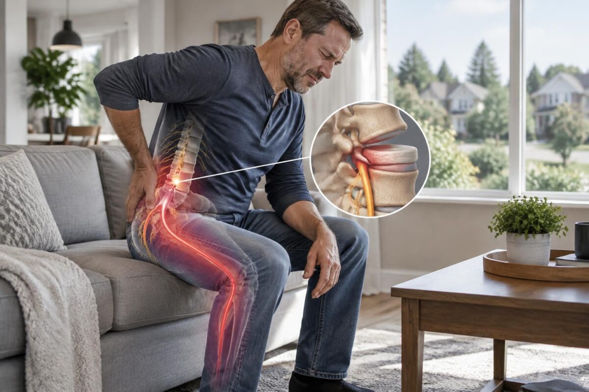
IN BRIEF Sciatica refers to pain that follows the path of the nerve in the buttock and leg; a herniated disc can irritate it, but an anomaly on the MRI is not sufficient to establish the cause. In Terrebonne, the…
Reading the words of the MRI report
Herniation, bulging, and disk degeneration
A report may mention disk bulging, protrusion, herniation, or disk degeneration. These terms describe changes observed on the image. They do not prove, alone, that the disk is the cause of the symptoms. Some individuals show changes on the MRI without pain, while others have marked symptoms with less visible abnormalities.
The radiologist may also describe contact or compression of a nerve root, as well as narrowing of the canal or a foramen. These observations make sense when compared with pain, sensitivity, strength, and reflexes. To understand the general steps for reading a lumbar MRI, refer to the guide to interpreting a lumbar spine MRI and this article on reading an MRI in the presence of a herniated disk.
Stenosis and disk pinching
The term stenosis refers to a narrowing of a space. Spinal stenosis concerns the central canal; foraminal stenosis concerns the opening through which a nerve root exits the spine. A report may also mention a decrease in disc height, sometimes referred to as disc compression. The professional evaluates whether these changes correspond to the location and type of symptoms.
An abnormal result does not automatically mean that intervention is necessary. To better understand common terms and possible follow-ups, you can read this article about an abnormal lumbar MRI and interpreting results. You can also consult this guide on the possible links between leg symptoms, disc bulges, and stenosis: lumbar MRI, disc bulges, stenosis, and leg symptoms.
Sciatica in Terrebonne: lumbar roots and the choice of a cautious treatment
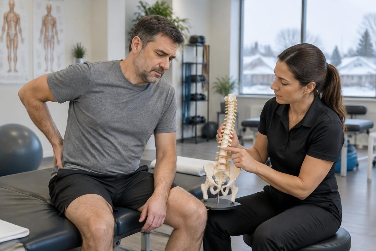
IN BRIEF Sciatica is pain related to the irritation or compression of a lumbar nerve root, which can radiate from the lower back to the buttock and leg. In Terrebonne, an evaluation allows for cautious treatment tailored to the cause…
Why the image must be put into context
An MRI is part of the evaluation, and not a complete diagnosis on its own. The healthcare professional compares the report to the symptoms, their evolution, and the results of the physical examination. This approach helps distinguish a visible anomaly that possibly explains the symptoms from an observation that is not directly related to the pain.
AI tools can assist in examining images or explaining certain terms, but their results do not replace the interpretation of a radiologist or the opinion of a licensed professional. It is also necessary to protect the confidentiality of medical information when sharing images or reports on an online platform. Any automated observation must be verified by a professional.

Sciatica in Terrebonne: how to interpret pain according to the leg path
IN BRIEF Sciatica is a pain that can start from the lower back or the buttock and travel down the leg to the foot. Its path, along with any tingling or weakness, helps guide the assessment but is not sufficient…
When to request an evaluation in Terrebonne
An evaluation can be useful if lower back pain persists, radiates into the leg, or is accompanied by changes in sensitivity or strength. Bring the report and, if you have them, the MRI images. The professional will examine the alignment between the symptoms and the mentioned levels, and then guide you based on your situation.
At the TAGMED Clinic, support options may include, depending on the evaluation, specific osteopathy or motorized neurovertebral decompression. These approaches do not replace a medical diagnosis. To learn more about services related to sciatica in Terrebonne, visit the clinic’s page.
Consult a doctor or emergency service promptly if you lose control of your bladder or bowels, experience numbness in the saddle area, have significant or progressive weakness of the leg or foot, fever after trauma, or a rapid worsening of pain.
About the author
Sylvain Desforges, B.Sc., D.O., N.D., osteopath and naturopath, is the founding president of TAGMED and the ACMA association. His practice focuses mainly on chronic pain of the spine, lower back pain, and symptoms compatible with sciatica, including when the pain radiates to the buttock, leg, calf, foot, or toes.
Through SOS Sciatique, he publishes educational content on sciatica symptoms, possible causes — such as disc herniation, disc compression, foraminal stenosis, or irritation of a lumbar nerve root — and non-surgical approaches that may be considered after an evaluation. Care at the TAGMED Clinic may include, as appropriate, motorized neurovertebral decompression, specific osteopathy, or precision percussion.
For an evaluation related to sciatica treatment in Montreal / Mont-Royal or sciatica treatment in Terrebonne, check the services available at the TAGMED Clinic.
Medical disclaimer
The information published on SOS Sciatique is provided for educational purposes. It does not replace a consultation, diagnosis, opinion, or treatment performed by a physician or another authorized healthcare professional. The author of this article is neither a doctor nor a specialist in a medical specialty recognized by the Collège des médecins du Québec. The osteopathic, manual medicine, or functional support approaches described on this site do not constitute medical acts and are not intended to replace medical follow-up. In case of loss of bladder or bowel control, numbness in the saddle area, significant or progressive weakness of the leg or foot, fever, recent trauma, severe pain, or rapid worsening, consult a physician or emergency service. For more details, please read our Legal notice.
Medical disclaimer: The information and advice provided on this site do not replace the advice, diagnosis, or treatment of a healthcare professional. Please note that Sylvain Desforges, B.Sc., D.O., N.D., Doctor of Osteopathy, is not a medical doctor and is not a specialist in a medical specialty as defined by the Collège des médecins du Québec. Manual medicine, functional medicine, and sports medicine as described on this site exclude any treatment or medical diagnosis made by a doctor or a medical specialist. Always consult your physician for any medical questions. For more details, please read our Complete Legal Notice.

Sciatica in Terrebonne: L5-S1, canal, foramen and nerve compression
IN BRIEF The sciatica L5-S1 corresponds to irritation or compression of a nerve root, sometimes related to a disc herniation or narrowing of the lumbar canal or the foramen. The pain may radiate from the lower back to the buttock,…
Sciatica in Terrebonne: identifying lumbar levels on an MRI
Upper levels
- L1-L2 and L2-L3: high lumbar levels; an impairment does not usually explain classic sciatica.
- L3-L4: may involve the L4 root and contribute to pain in the front of the thigh or the knee.
Levels often examined in cases of sciatica
- L4-L5: may irritate the L5 root; pain sometimes radiates to the leg and the top of the foot.
- L5-S1: may affect the S1 root; pain sometimes radiates to the calf and the outer edge of the foot.
- To remember: the image alone does not confirm the cause; symptoms and the clinical examination matter.

In Terrebonne, understanding the lumbar levels mentioned in an MRI can help better discuss sciatica with a healthcare professional. The levels L1 to S1 describe the location of the vertebrae and discs; however, an anomaly visible on the image does not prove, on its own, that it causes your symptoms. The interpretation must take into account pain, the clinical examination, and the radiologist’s report.
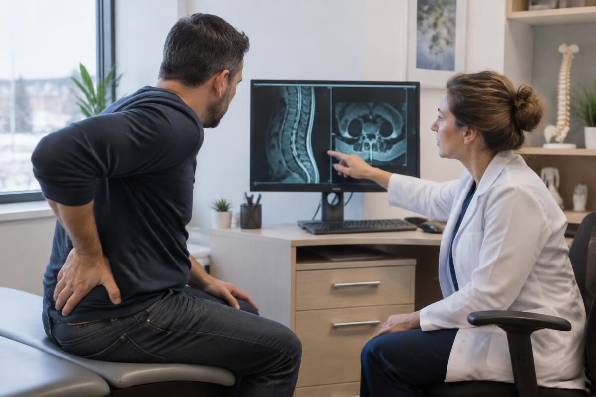
Sciatica in Terrebonne: why an MRI must be correlated with symptoms
IN BRIEF Sciatica is a pain that often radiates from the lower back to the buttock and leg. A lumbar MRI shows the structures of the spine, but its results must correspond to the symptoms and clinical examination: a visible…
Identifying lumbar levels on the MRI report
What do L1 to L5 and S1 mean?
The lumbar region includes five vertebrae, designated from L1 to L5. Below L5 is the sacrum, the upper part of which is called S1. The discs are named according to the two vertebrae they separate: the L4-L5 disc is located between L4 and L5, while L5-S1 is found between L5 and S1.
On the images, the levels can be identified by following the column from top to bottom. The report typically uses these landmarks to describe a protrusion or herniated disc, spinal canal narrowing, or a decrease in space through which a nerve root passes. The radiologist also identifies the affected side, right or left, and sometimes specifies if the change is located centrally or near a foramen.
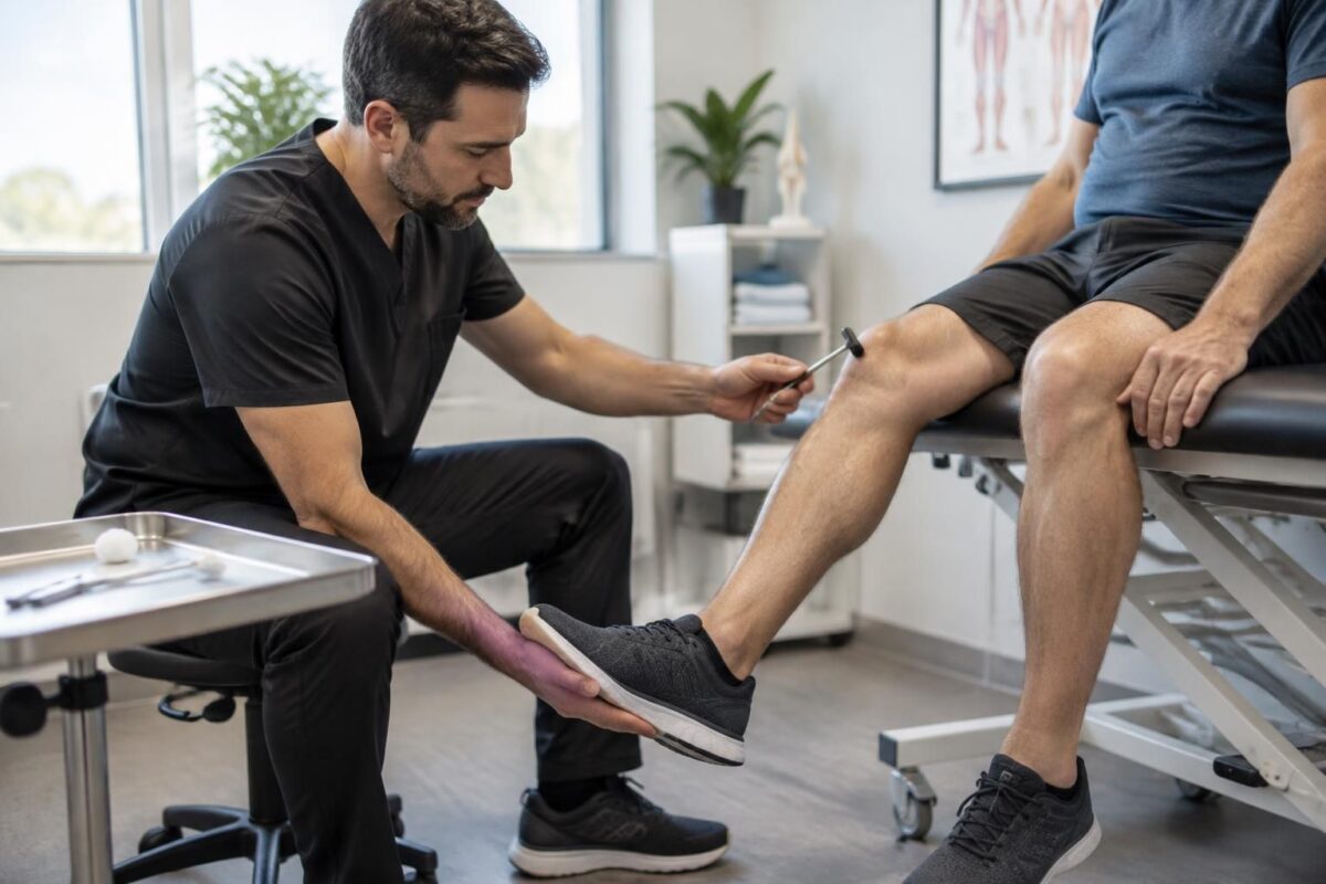
Sciatica in Terrebonne: reflexes, strength, and sensitivity in evaluation
IN BRIEF In Terrebonne, the assessment of sciatica checks the reflexes, strength, and sensitivity of the legs to identify possible nerve damage. These observations, combined with the pain’s pathway and its evolution, help to guide the next steps and recognize…
Linking observations to sciatica symptoms
Nerve irritation can lead to pain that radiates from the buttock to the thigh, calf, or foot. Certain levels, particularly L4-L5 and L5-S1, are frequently mentioned in examinations of individuals presenting with sciatica symptoms. However, the location of the pain does not always allow for precise determination of the affected level. Sensations of numbness, tingling, or weakness may also guide the evaluation, but do not replace a professional examination.
It is recommended to read the report with a physician or a licensed healthcare professional. Ask them to clarify which level is described, which nerve might be involved, and if this observation corresponds to your symptoms and the findings from the physical examination. Avoid concluding that a herniated disc is the certain cause of your pain based on an isolated image: disc changes may be present in individuals without pain.
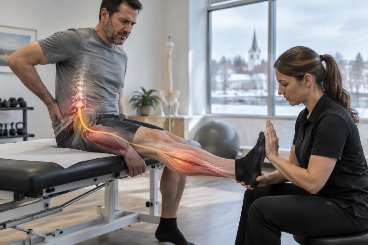
Sciatica in Terrebonne: muscle weakness according to the irritated nerve root
IN BRIEF Sciatica with muscle weakness can occur when a lumbar nerve root is irritated; affected movements, such as raising the foot or walking on tiptoes, provide clues but are not sufficient for a diagnosis. In Terrebonne, an evaluation can…
Practical tips before and after the exam
Bring the complete report and, if possible, the MRI images to your appointment. Note how long the symptoms have been occurring, their pathway, the movements that aggravate or relieve them, and any changes in strength or sensitivity. This information helps contextualize the imaging results within your situation.
An MRI is not necessary for every episode of lower back pain. The professional assessing you will decide if it is indicated based on the evolution of symptoms and the observed signs. In Terrebonne, a clinical assessment can help discuss next steps and non-surgical support options when appropriate. Manual therapies do not replace a medical diagnosis or recommended follow-up.
When to seek urgent help
Consult a physician or emergency service quickly in cases of loss of urinary or bowel control, numbness in the saddle area, significant or progressive weakness of the leg or foot, fever, recent trauma, severe pain, or rapid worsening.
About the author
Sylvain Desforges, B.Sc., D.O., N.D., osteopath and naturopath, is the founding president of TAGMED and the ACMA association. His practice focuses particularly on chronic spinal pain and pain consistent with sciatica.
Medical disclaimer
This content is provided for educational purposes and does not replace a consultation, diagnosis, or treatment by a licensed healthcare professional. The author is neither a physician nor a specialist in a medical specialty recognized by the Collège des médecins du Québec. Osteopathy and manual medicine approaches do not replace medical follow-up.
Landmarks of lumbar levels on an MRI
| Observed level | Possible marker for sciatica |
|---|---|
| L3-L4 | Nerve irritation may contribute to pain in the front of the thigh or knee. |
| L4-L5 | Affecting the L5 root may be associated with pain radiating to the side of the leg and the top of the foot. |
| L5-S1 | Irritation of S1 may correspond to pain behind the leg, extending to the edge of the foot. |
| Herniated disc | Its position and contact with a nerve root matter more than its mere presence. |
| foraminal stenosis | Narrowing near a root may be relevant if it matches the symptoms. |
| Clinical reading | The level visible in the MRI does not confirm the origin of the pain on its own. |
Illustrative testimonials in Terrebonne: understanding lumbar levels on an MRI
These testimonials are fictional and serve only to illustrate common questions. They do not represent real patient opinions and do not allow for a diagnosis to be made. The interpretation of an MRI must be performed by a qualified healthcare professional, taking into account the symptoms and the clinical examination.
“My report mentioned L4-L5, but I didn’t know what that meant.” In this example, a person from Terrebonne describes pain that starts in the lower back and radiates to the leg. The references L1 to L5 denote the lumbar vertebrae; mentions like L4-L5 indicate the space between two vertebrae, where a disc is located. A change observed at this level does not prove, on its own, that it is the cause of the pain.
“I was told about L5-S1 and I thought that necessarily indicated sciatica.” L5-S1 corresponds to the junction between the last lumbar vertebra and the sacrum. Irritation of a nerve root in this area can sometimes be associated with pain that radiates down into the buttock or leg. However, the distribution of symptoms varies and must be compared to the results of the medical examination and imaging.
“My MRI showed a disc protrusion, but my symptoms didn’t exactly match what I was reading.” This scenario serves as a reminder that an MRI image must be interpreted with caution. Disc peculiarities may be present without causing symptoms. The professional evaluates their location, the neighboring nerve structures, and the correlation with pain, numbness, or possible weakness.
“I wanted to understand if the level mentioned in my report explained my foot pain.” Some lumbar nerve roots contribute to the sensation and strength of different parts of the leg and foot. But the territories overlap, and symptoms do not always follow a perfectly clear pattern. It is therefore better to have the report explained in its entirety rather than relying on a single term or image.
During an evaluation for sciatica in Terrebonne, it may be helpful to bring the MRI report and, if possible, the images themselves. A discussion can help clarify terms such as “disc herniation,” “foraminal narrowing,” or “contact with a nerve root,” without presuming that each finding necessarily explains the symptoms.
Consult a doctor or emergency service quickly in case of urinary or bowel incontinence, numbness in the saddle region, significant or progressive weakness in the leg or foot, or rapid worsening. An MRI and this educational information do not replace a medical evaluation.
Sylvain Desforges, B.Sc., D.O., N.D., osteopath and naturopath, is the founding president of TAGMED clinics and the ACMA association. His practice focuses, in particular, on chronic spinal pain, lower back pain, and symptoms compatible with sciatica. He is also interested in the thoughtful integration of technologies such as neurovertebral decompression, laser, and shockwaves, as part of an approach tailored to each person’s situation.
Through his educational content, Sylvain Desforges aims to help readers better understand the examinations and approaches that can be discussed during an evaluation. The theme of sciatica in Terrebonne often raises questions about the MRI: what do the lumbar levels represent, and how can an image be related to pain that radiates down into the buttock or leg? The MRI provides detailed images of discs, soft tissues, and nerve structures, but by itself, it is not sufficient to establish the cause of symptoms.
The lumbar region consists of five vertebrae, designated from L1 to L5. Between most of them lie discs, named according to the vertebrae that frame them: for example, the L4-L5 disc is located between the fourth and fifth lumbar vertebrae. Below L5 is the L5-S1 level, at the junction between the lumbar spine and the sacrum. These landmarks allow for precise descriptions of where a report mentions a disc protrusion, herniation, or narrowing.
Irritation or compression of a nerve root can sometimes contribute to sciatica-like symptoms. Depending on the affected level, pain, tingling, or numbness may manifest in different areas of the leg or foot. However, anatomical descriptions do not automatically determine the source of a symptom: clinical examination, the history of pain, and other results must also be considered. Moreover, some abnormalities visible on MRI may be present in individuals who do not have pain.
Understanding the terms of a report can therefore help prepare for a discussion with a healthcare professional, without replacing their interpretation. An evaluation may consider strength, sensitivity, reflexes, movements that alter symptoms, and their progression. An MRI is generally considered in context, particularly when symptoms persist, when there are neurological signs, or when imaging is necessary to clarify the subsequent management.
At TAGMED Clinic, care options are determined based on the evaluation and reported needs; they may include, when appropriate, motorized neurovertebral decompression, specific osteopathy, or precision impactor. These approaches do not replace a medical diagnosis or follow-up with a licensed healthcare professional. A consultation related to sciatica treatment in Terrebonne allows for discussion of the available services and their relevance to the individual situation.
Medical warning: this information is educational and does not replace a consultation, diagnosis, or treatment by a physician or other licensed healthcare professional. The author is neither a doctor nor a specialist in a medical specialty recognized by the Collège des médecins du Québec. Consult a doctor or emergency service quickly in case of urinary or bowel incontinence, anesthesia in the saddle region, significant or progressive weakness, fever, recent trauma, or rapid worsening.
Understanding the lumbar levels indicated on an MRI helps to locate the discs, nerves, and changes described in the report when pain suggests sciatica in Terrebonne. Mentions like L4-L5 or L5-S1 refer to specific levels in the lower back, but a visible abnormality on the image is not sufficient, by itself, to explain symptoms: interpretation must take into account their location, progression, and clinical examination.
Markers to Understand Lumbar Levels
The lumbar spine consists of five vertebrae, named from L1 to L5. Between them are discs, and below L5 is the sacrum. A level such as L4-L5 corresponds to the space between the fourth and fifth lumbar vertebrae; L5-S1 designates the junction between the last lumbar vertebra and the sacrum. These markers allow the professional to locate a disc protrusion, herniation, or narrowing observed on the MRI.
The nerve roots exit the spine at different levels and contribute to sensitivity and strength in the leg and foot. However, the pathways of symptoms vary from person to person and may overlap. Foot pain that worsens in a seated position, for example, deserves to be precisely described to the professional; it alone does not determine which level is involved. You can refer to this page on foot pain worsened in a seated position as well as this article on deep buttock pain radiating into the thigh.
What the MRI can show — and what it does not confirm alone
The MRI provides detailed images of the discs, soft tissues, spinal canal, and nerve structures. The report may describe disc pathology, protrusion, herniation, foraminal narrowing, or lumbar stenosis. To understand some of these terms, check the resources on disc pathology, protrusion, and herniation in lumbar MRI and on lumbar stenosis and its possible links to sciatica.
An anomaly may be present without causing pain, while marked symptoms do not always correspond to a spectacular image. The key question is whether the described level and side correspond with the symptoms and clinical examination results. Additional explanations on interpreting a report are available in this guide to understanding a back MRI report and in this article on signs such as black disc, protrusions, and Modic changes, which do not necessarily warrant premature alarm.
Connecting images to leg symptoms
To prepare for a consultation, note where the pain starts, how far it descends, the positions that worsen it, and the possible presence of tingling, numbness, or weakness. Numbness that progresses towards the foot or a change in strength should be reported quickly to the healthcare professional. For more depth on these topics, see the pages dedicated to leg numbness, numbness that advances towards the foot, and sciatica and its possible manifestations.
Pain that makes long trips difficult, such as increased pain while driving, is also useful information to report. Describing the tolerated duration in a seated position and changes in symptoms helps complete the interpretation of the images; consult this resource on pain that prevents long driving for more on this.
Interpretation and next steps
An MRI must be interpreted by a qualified professional, taking into account the report, symptoms, and clinical examination. Automated tools or image analysis systems may provide general information, but they do not replace a diagnosis or a consultation. Bring your images and the report to your appointment, and ask which level is involved, if a nerve root appears to be compressed, and how the observations align with your symptoms.
In the case of loss of bladder or bowel control, anesthesia in the saddle area, or significant or progressive weakness in the leg or foot, seek urgent medical evaluation without delay. These signs require special attention and should not be evaluated solely based on an MRI or online information.

Sylvain Desforges, B.Sc., D.O., N.D., docteur en ostéopathie
Dr Sylvain Desforges, B.Sc., D.O., N.D., osteopath, is the founding president of TAGMED and of the ACMA association. He dedicates his practice to chronic spinal pain, low back pain and symptoms compatible with sciatica, particularly when pain radiates toward the buttock, leg, calf, foot or toes.
Through SOS Sciatique, he publishes educational content on sciatica symptoms, possible causes such as disc herniation, disc narrowing, foraminal stenosis or irritation of a lumbar nerve root, as well as non-surgical approaches that may be considered according to the evaluation. At Clinique TAGMED, care may include, depending on the case, motorized neurovertebral decompression, specific osteopathic care or precision percussion.
For an evaluation related to sciatica treatment in Montreal / Mount Royal or sciatica treatment in Terrebonne, please consult the services available at Clinique TAGMED.
Medical disclaimer
The information published on SOS Sciatique is provided for educational purposes only and does not replace a consultation, diagnosis, opinion or treatment provided by a physician or another authorized healthcare professional. The author of this article is neither a medical doctor nor a specialist in a medical specialty recognized by the Collège des médecins du Québec. The osteopathic, manual medicine or functional support approaches described on this site do not constitute medical acts and are not intended to replace medical follow-up.
Consult a physician or emergency service in cases of loss of bladder or bowel control, anesthesia in the saddle area, significant or progressive weakness in the leg or foot, fever, recent trauma, severe pain or rapid worsening of symptoms. For more details, please read our Legal Notice.