




<!–
DEDICATED PAGE — SOS SCIATIQUE TOOL 03
Editorial title: Test L4, L5 or S1: locate the path of the pain
Slug: test-l5-s1-sciatic-path
Canonical URL: https://www.sossciatique.com/test-l5-s1-trajet-sciatique
Featured image: https://www.sossciatique.com/wp-content/uploads/2026/07/test-l5-s1-trajet-sciatique-hero.png
Page images:
– https://www.sossciatique.com/wp-content/uploads/2026/07/test-l5-s1-trajet-sciatique-hero.png
– https://www.sossciatique.com/wp-content/uploads/2026/07/test-l5-s1-trajet-sciatique-l5.png
– https://www.sossciatique.com/wp-content/uploads/2026/07/test-l5-s1-trajet-sciatique-s1.png
– https://www.sossciatique.com/wp-content/uploads/2026/07/test-l5-s1-trajet-sciatique-evaluation.png
– https://www.sossciatique.com/wp-content/uploads/2026/07/test-l5-s1-trajet-sciatique-cta.png
CSS: no CSS is included on this page. It uses the already installed global CSS SOS Sciatique.
no script is included on the page. Activate the snippet Code Snippets "SOS Sciatique — Test L4 L5 S1" on the public part of the site.
RANK MATH — FRENCH
SEO title: Test L4, L5 or S1: path of the sciatic pain
Meta description: Where does your pain go? This L4, L5, or S1 test compares path, numbness, and weakness to provide an educational profile, without diagnosis.
Focus Keywords: test L5 S1, pain L5 or S1, sciatic path, sciatic nerve root, sciatic foot pain
Tags: sciatica, L4, L5, S1, sciatic test, pain path, nerve root, leg pain, foot pain, cruralgia, radiculopathy
RANK MATH — ENGLISH
SEO Title : L4, L5 or S1 test: map your sciatica pain
Meta Description : Where does your pain travel? This L4, L5 or S1 test compares pain, numbness and weakness patterns to give educational guidance, not a diagnosis.
Focus Keywords : L5 S1 test, L5 or S1 symptoms, sciatica pain map, nerve root pain, sciatica foot pain
Tags : sciatica, L4, L5, S1, sciatica test, pain pattern, nerve root, leg pain, foot pain, radiculopathy, femoral nerve pain
–>
Test L4, L5 or S1: locate the path of the pain
Does your pain travel down the front of the thigh, along the side of the leg, or behind the calf? This questionnaire compares the painful path, areas of numbness, and movement difficulties often associated with the L4, L5, and S1 roots.
The result indicates the most compatible profile with your responses. It does not confirm a compressed root, a herniated disc, or the cause of your symptoms.
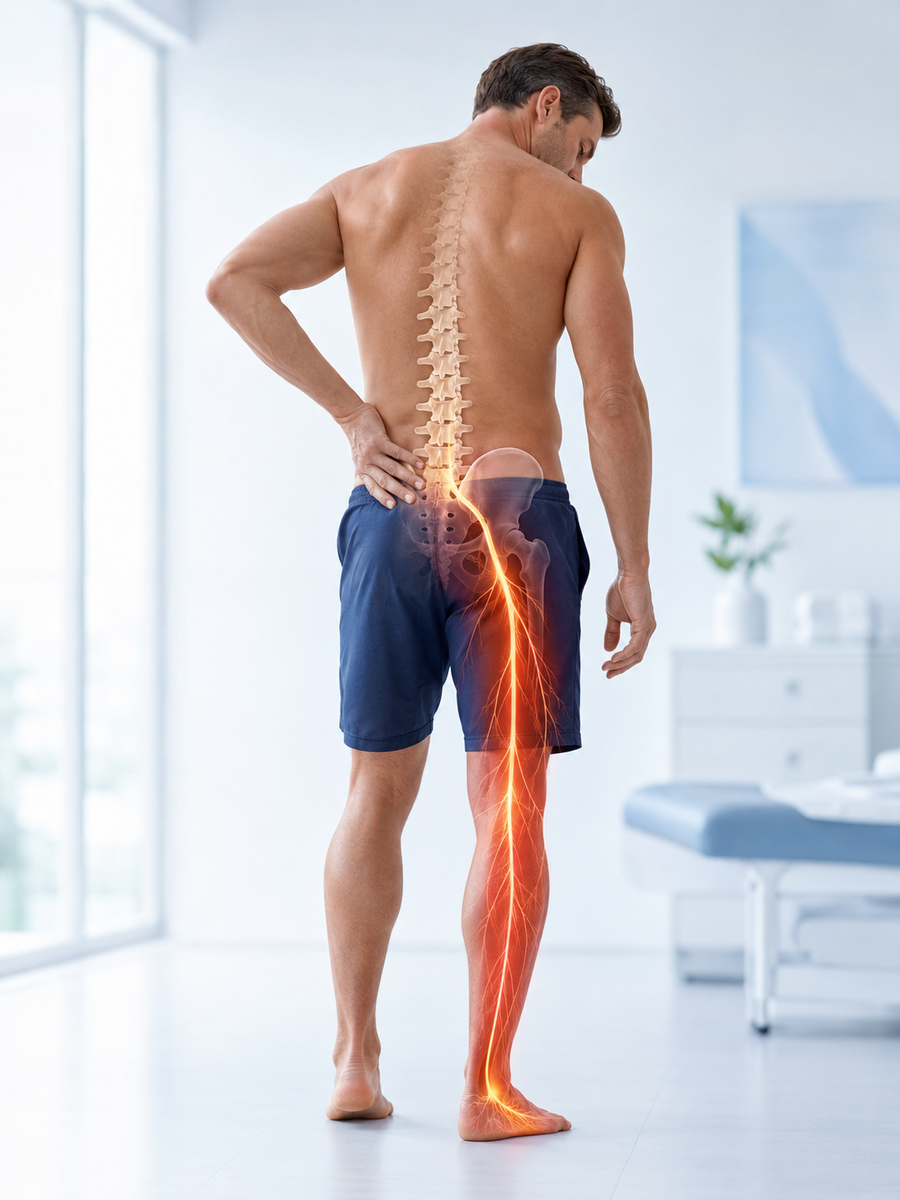
Which route resembles your symptoms the most: L4, L5, or S1?
Answer the eight questions based on your current situation. The tool prioritizes signs that require urgent consultation.
Question 1 of 8
Please choose an answer before continuing.
Symptoms Map for L4, L5, and S1
No skin map perfectly corresponds to all individuals. Research indicates a significant overlap of painful radicular pathways. The table below is therefore intended to organize clues, rather than serve as a self-diagnosis tool.
| Root | Pain or sensitivity often described | Occasionally weakened movement | Reflex sometimes diminished |
|---|---|---|---|
| L4 | Front of the thigh, knee, inner side of the leg or ankle | Knee extension | Patellar |
| L5 | Side of the leg, top of the foot, big toe | Foot or big toe lift | No reliable single reflex |
| S1 | Back of the calf, heel, sole or outer edge of the foot | Plantar flexion, rising on tiptoes | Achilles |
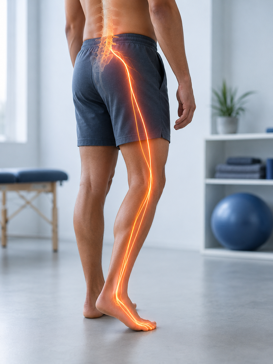
What symptoms are more indicative of the L5 root?
The L5 pain may start from the buttock, run along the outer thigh or leg, and then reach the top of the foot and the big toe. Tingling or numbness may cover part of this same area. The pain does not need to follow the entire line to be clinically relevant.
In terms of motor function, L5 involvement can make it more difficult to lift the foot or the big toe. A person may trip on their toes, feel less control when walking on their heels, or, in more marked cases, present with foot drop. This finding warrants a professional assessment; if the weakness is recent or worsening, advice should be sought promptly.
Does L4–L5 always refer to L5?
A disc herniation located between L4 and L5 often affects the L5 root descending in that area. However, a very lateral herniation may impact the L4 root exiting at the same level. Thus, reading an imaging report should be correlated with symptoms and examination findings.
What symptoms are more indicative of the S1 root?
The S1 pathway is often posterior: buttock, back of the thigh, calf, heel, sole, or outer edge of the foot. Abnormal sensations may affect the small toes. For some individuals, the pain is particularly concentrated in the calf or foot rather than throughout the entire limb.
S1 contributes to the downward push of the foot. Therefore, involvement may make it more difficult to rise on tiptoes or repeatedly lift the heel. The Achilles reflex may be diminished, but this verification belongs to the clinical examination.
Does L5–S1 always refer to S1?
A herniated disc L5–S1 located in the most common position often irritates the S1 root. As with L4–L5, the exact location of the lesion and individual variations can change the affected root. An image alone does not prove that the observed lesion explains the pain.
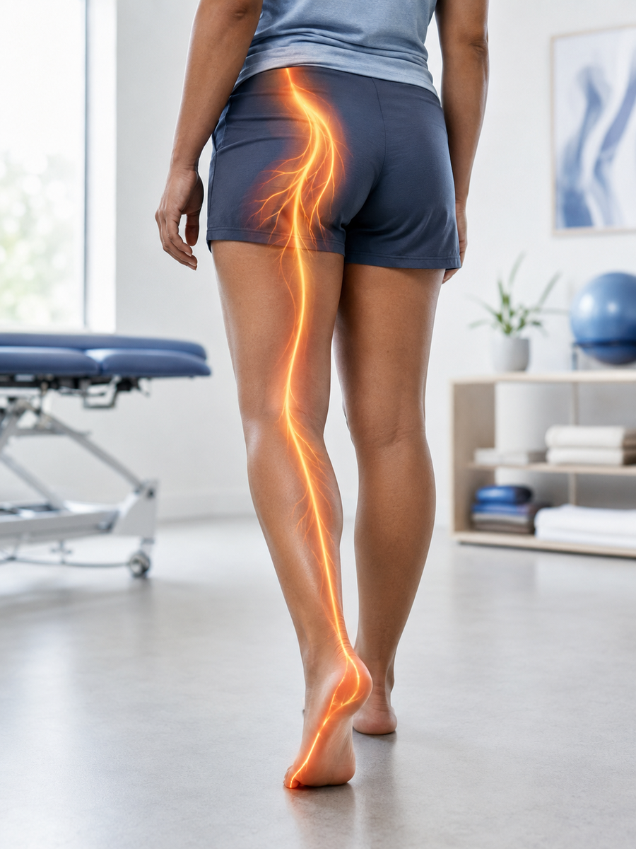

How does a professional verify the affected root?
The interview clarifies the onset of symptoms, their progression, the exact path, the presence of nerve sensations, and the effect on walking or activities. The examination can then compare the strength of specific muscle groups, the sensitivity of different areas, reflexes, mobility of the spine and hip, as well as some nerve tension tests.
A single element is rarely decisive. For example, difficulty in raising the foot may stem from L5, but also from a peripheral nerve or another condition. The interpretation becomes stronger when the pathway, sensitivity, strength, and reflexes converge.
MRI is not automatically required for every radicular pain. It becomes more relevant when there is a neurological deficit present, when warning signs exist, when the evolution is unusual, or when the result is likely to alter treatment.
How is the result of the L4, L5, or S1 test calculated?
The algorithm assigns separate points to the L4, L5, and S1 profiles. The main pathway receives the highest weight, followed by the area of numbness, the type of weakness, and the lowest point reached by the pain. The predominance of pain in the leg, its unilateral nature, and its increase with coughing or exertion serve as secondary clues to radicular irritation.
A specific profile is displayed only if one territory sufficiently dominates the other two. When responses overlap, are contradictory, or lack nerve clues, the result is classified as ‘mixed or indeterminate’. Warning signs and progressive weakness always replace the anatomical result with a safety orientation.
Frequently Asked Questions about L4, L5, and S1 Pain
Can this test confirm that my pain comes from L5 or S1?
No. It compares your responses to common profiles. The localization of a root requires a clinical examination and, in some cases, imaging or other tests.
What is the difference between L5 sciatica and S1 sciatica?
L5 often manifests on the side of the leg, the top of the foot, and the big toe, sometimes with difficulty lifting the foot. S1 more often goes behind the calf towards the heel, the sole, or the outer edge of the foot, sometimes causing difficulty on tiptoe.
Is L4 pain sciatica?
The L4 root mainly contributes to the femoral nerve. L4 pain located in the front of the thigh is therefore generally called cruralgia or L4 radiculopathy rather than sciatica in the strict sense.
Does an L4–L5 herniation affect the L4 or L5 root?
In its most common position, it affects the L5 root that passes through this level. A foraminal or very lateral herniation may rather reach L4. The imaging report must be interpreted alongside the examination.
Does an L5–S1 herniation always affect S1?
It often affects S1, but not always. The position of the herniation, individual anatomy, and other lesions can modify the affected territory.
Can there be L5 damage without pain in the big toe?
Yes. The pathways can be partial, vary over time, or overlap. The absence of a classic symptom does not rule out damage, just as its presence does not confirm it.
Why do my symptoms correspond to both L5 and S1?
The sensory territories overlap, the description of the pain varies, and more than one root can sometimes be irritated. A non-radicular referred pain can also blur the profile.
Can I test my strength by walking on my heels or toes?
These movements sometimes provide a clue, but they should not be forced in the presence of pain, imbalance, or fall risk. A professional can compare strength in a safer and more precise manner.
When does foot weakness become urgent?
A foot drop, new weakness, rapid worsening, or a leg giving way justifies prompt medical advice. If it is accompanied by urinary, bowel problems, or numbness in the perineum, evaluation should be immediate.
Do I need an MRI to know if it’s L5 or S1?
Not necessarily. A clinical examination can often guide the territory. Imaging is discussed when its results may change the management, in the presence of a deficit or when the evolution is concerning or atypical.
Does foot numbness prove a herniated disc?
No. A lumbar root may be involved, but a peripheral nerve, neuropathy, or another condition can produce similar numbness.
What should I do if my result is mixed or indeterminate?
Monitor the evolution without ignoring warning signs. Consult if symptoms persist, progress, limit your activities, or if you have weakness or decreased sensitivity.
Clinical Sources and References
- Health Insurance — Sciatica, how to recognize it?
- Health Insurance — Consultation and treatment for sciatica
- StatPearls — Lumbar Disc Herniation
- StatPearls — Lumbosacral Radiculopathy
- Albert et al. — Accuracy of pain drawings in identifying the involved nerve root
- NICE — Low back pain and sciatica in over 16s
Sylvain Desforges, B.Sc., D.O., N.D., Doctor of Osteopathy
The information on this page is educational. It does not replace the advice, diagnosis, or treatment of a physician or another qualified healthcare professional.
Your journey seems compatible with L4, L5 or S1?
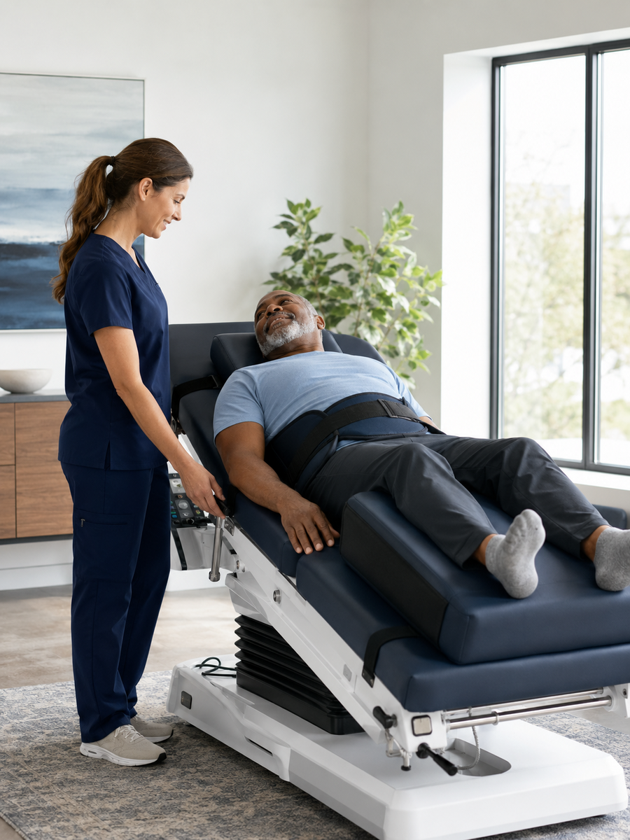
A planned evaluation can compare pain areas, check strength and sensitivity, examine the back, hip, and peripheral nerves, then determine if a nonsurgical approach is relevant.
If the result indicates an emergency or urgent medical advice, follow this recommendation rather than scheduling a regular appointment.
Sylvain Desforges, B.Sc., D.O., N.D., Doctor of Osteopathy
Editorial information, sources and limitations
This content is intended to inform patients about sciatica, possible causes, warning signs, and care options. It does not replace an individualized assessment.
Reference sources
References are selected according to the subject of the page: guidelines, systematic reviews, then institutional resources.
- NCBI Bookshelf – Sciatica — Clinical institutional resource
- AAOS OrthoInfo – Sciatica — Patient resource from a professional society
- ameli – Sciatica — French institutional patient resource
Complementary resources from the TAGMED network
These internal resources complement the clinical information and thematic linking. They do not replace national guidelines or systematic reviews.
Limitations of this information
The information on this page is general. It does not constitute a diagnosis, prescription, or guarantee of results. Pain radiating into the leg may have several causes; assessment should consider clinical history, examination findings, symptom progression, and, when appropriate, complementary tests.
When to seek urgent medical care
Seek urgent medical care if you experience loss of bladder or bowel control, saddle anesthesia, major or progressive leg weakness, unexplained fever, pain after significant trauma, or severe pain that rapidly worsens.