

Terrebonne sciatic treatment: what to do when the pain descends into the leg?
30/06/2026
Sciatica: why the cause must guide the choice of treatment
30/06/2026
In short In short, heel pain (Talalgia) may be due to causes locals or root damage such as Root S1. This distinction allows a rapid diagnosis and an Appropriate support. non-surgical approaches are based on drill, of braces and manual techniques. How does it work? Targeted evaluation, differentiation of causes and personalized treatment plan. In Montreal, this method is particularly recommended for quick and lasting results, with pain reduction and improvement in walking. |
Heel pain: S1 or other cause?
- main reason : heel pain is most often of local origin (plantar fasciitis, calcaneal spine, Achilles tendinitis, calcanean bursitis), but it can also come from a S1 radiculopathy When the pain follows a path from the lower back to the foot and is accompanied by neurological signs.
- Signs of S1 radiculopathy : pain that extends from the lumbar to the calf and foot, possible weakness of the foot (tissue), decrease in the reflexachillé, little local improvement at rest.
- Signs of a local cause : Pain localized under the heel, aggravated in support on hard ground or on waking, pain on palpation near the heel, swelling or stiffness of adjacent tissues.
- When to consult : severe pain preventing any support, significant numbness, progressive weakness, fever, recent trauma, or persistence beyond 2 weeks despite personal measures.
- Quick measurements : 15–20 minutes glaze, relative rest, foot elevation, light compression, stretching of the plantar fascia and calves, suitable shoes and, if necessary, orthoses.
Quick Summary: This article explains the possible causes of heel pain, distinguishes pain related to an S1 root from other local origins, and indicates how to quickly assess and take care of these situations. You will understand the signs that are directing towards a common local cause and those that suggest a nervous conflict. The text offers practical options at the TagMed Clinic and emphasizes when to consult for a reliable diagnosis.
Frequent local causes of the heel
plantar fasciitis. It is an inflammation of the plantar fascia, a band that connects the heel to the toes. It affects a large part of the population and often occurs after repeated overload. The pain occurs when you wake up and can burn or pull the underside of your foot when you start. It can improve briefly and then come back at the end of the day. The diagnosis is based on observing the painful motive and clinical examination.
calcaneal thorn. This bone protrusion under the heel can cause targeted pain in the center of the heel, especially on hard surfaces. It can be asymptomatic in some people. Treatments include pads, orthopedic insoles, icing and, if necessary, targeted therapies in the clinic.
Achilles tendinitis. The inflammation of the tendon connecting the calf to the heel is manifested by posterior pain in the heel, sometimes accompanied by swelling and morning stiffness. The use of progressive stretching, heel pads and adaptation of activity can help, while avoiding sudden movements and direct shocks in the region.
Calcanean bursitis. Inflammation of the purse around the heel due to rubbing or an unsuitable shoe. Localized pain, swelling and heat on the back of the heel. The temporary stopping of rigid shoes, icing and local protection are part of the treatment.
More rarely, other local causes exist, such as fatigue fractures or inflammatory problems. A consultation allows you to specify the diagnosis and to adapt the treatment.
Pain of root root S1: signs and context
Pain reflecting participation of the lumbar nerve roots may affect the heel and sole of the foot. The key sign is the pain that follows the path of the sciatic nerve, often associated with pain in the lower back or in the leg. Tingling, weak foot thrust or decreased Achilles reflex can accompany the picture.
S1 root pain is not limited to the heel and may be accompanied by lumbar pain or low back pain radiating to the feet. Strictly localized pain in the heel without any other neurological sign may instead point to a local cause. If neurological signs appear, a clinical examination and, if necessary, imaging is recommended.
Diagnosis and orientation
The diagnosis is based on history, clinical examination and comparison of painful reasons. In the presence of marked neurological symptoms, noticeable weakness or painful trafficking following a dermatosome, specialized treatment is indicated. In some cases, additional examinations will be considered to assess the state of the nervous and rhabdom structures, while respecting the general medical recommendations.
To understand the links between sciatica and heel pain and explore specialized opinions, you can consult dedicated resources on the following areas: sossciatica.com/pain-to-calf-and-sciatica, SoshernieDiscale.com, and SosherniateDisc.com.
Indicative diagnosis and management
An evaluation by a health professional makes it possible to distinguish a local cause from a radiculopathy. In case of suspicion of a disc process or neurovertebral decompression, the recommendations favor a suitable approach and avoid inappropriate exercises. The TagMed Clinic can propose non-surgical approaches adapted to each situation and direct to the relevant resources when necessary.
To deepen the options available in Montreal or Terrebonne, you can consult the resources of Tagmed Clinic and specialized resources on disc issues when needed via sites SoshernieDiscale.com and SosherniateDisc.com.
Support options and practical tips
In case of localized heel pain, simple measures can help: Adapted shoes with good heel support, Plantar orthoses To redistribute pressure and support the arch, cushions or heel pads And, when necessary, use of ice cubes to reduce inflammation. Specific protections and protections of the painful area can also be considered depending on the context.
Neurovertebral decompression and disc problems, when present, require a specialized assessment. In these situations, general recommendations avoid stretching or inappropriate exercise programs and favor targeted medical follow-up.
For additional information on the correspondence between heel pain and associated pathologies, you can consult decompressionneurovertebrale.com, as well as specialized resources on associated conditions in osteopathy and manual medicine.
Medical disclaimer
Medical Disclaimer: The information and advice provided on this site does not replace the health professional’s diagnosis or treatment. Please note that Dr. Sylvain Desforges osteopath is neither a doctor of medicine nor a doctor, and is not a specialist in a medical specialty as defined by the Collège des Médecins du Québec. Manual medicine, functional medicine and sports medicine as described on this site exclude any medical treatment or diagnosis made by a doctor or specialist doctor. Always consult your doctor for any medical questions. For more details, please read our full legal notice.
Looking for a specialized consultation? Request an assessment from Clinique TagMed or explore the following resources for orientations related to similar contexts: Tagmed Clinic, sossciatica.com/pain-to-calf-and-sciatica, sossciatica.com/sciatica-and-pinch-discal, and resources on herniated discs via SoshernieDiscale.com or SosherniateDisc.com.
Diagnostic axis : Nervous origin (S1) vs local causes in the heel.
- Root S1 : Pain following the nerve path to the heel.
- Other nervous causes : Lumbar irritation without obvious radiculopathy.
- plantar fasciitis : inflammation of the plantar fascia, morning pain.
- calcaneal thorn : protrusion under the heel, central pain on hard surfaces.
- Achilles tendinopathy : posterior heel pain, repeated overload.
- Calcaneal bursitis : inflammation of the purse, pain in the back of the heel.

This article helps you distinguish heel pain linked to an S1 root from a local heel cause, describing key signs, simple tests and management options to direct the most suitable treatment quickly.
Heel pain and S1 root: how to recognize nervous involvement
S1 radiculopathy May be manifested by pain that follows the path of the nerve S1, with irradiations towards the calf and the outer edge of the foot. You may feel weakness in plantar flexion (foot tip), decreased tendon reflection and sometimes sensory disturbances on the lateral side of the foot. This pain can be accompanied by postural disarray and mild to moderate lameness when the spine is irritated. An accessible examination may show increased pain in nerve stretching or some foot and ankle movement tests. In the presence of these signs, a medical assessment should be considered to exclude more general causes and specify the diagnosis.
Signs that direct to a local cause of the heel
The Local heel pain is often centered on the heel itself and worsens with walking, hard surfaces or when rising in the morning. Among the most frequent causes are the plantar fasciitis, thecalcaneal thorn, the Achilles tendinitis and the Calcaneal bursitis. In plantar fasciitis, the pain is typically more marked on waking and briefly subsides after a few steps before returning. The calcaneal spine can cause central and acute pain when walking on a hard area. Achilles tendonitis is manifested by pain in the back of the heel with possible morning stiffness and swelling. Bursitis is characterized by localized pain, heat or swelling at the back or at the top of the heel. Each scenario has its particularities and requires a suitable approach.
How to differentiate and guide care
To distinguish S1 radiculopathy from a local cause, it is necessary to consider the extent of the pain (radiating towards the foot and the posterior portion of the leg vs strictly localized at the heel), motor signs and simple examinations. In case of suspicion S1, targeted imaging and specialized consultation may be indicated, especially if neurological symptoms progress or are accompanied by loss of urinary control, significant numbness or marked weakness. In the presence of strictly heel pain without root irradiation, the treatment generally aims at local relief and progressive strengthening of the support structures of the foot.
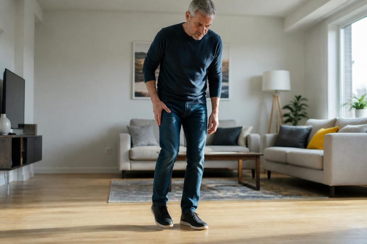
Sciatica and foot weakness: risk of drop foot
IN BRIEF In brief, sciatica and weakness of the foot can cause a drop foot, the inability to lift the front of the foot. This situation occurs when the peroneal nerve or the sciatic nerve is compressed or injured. The…
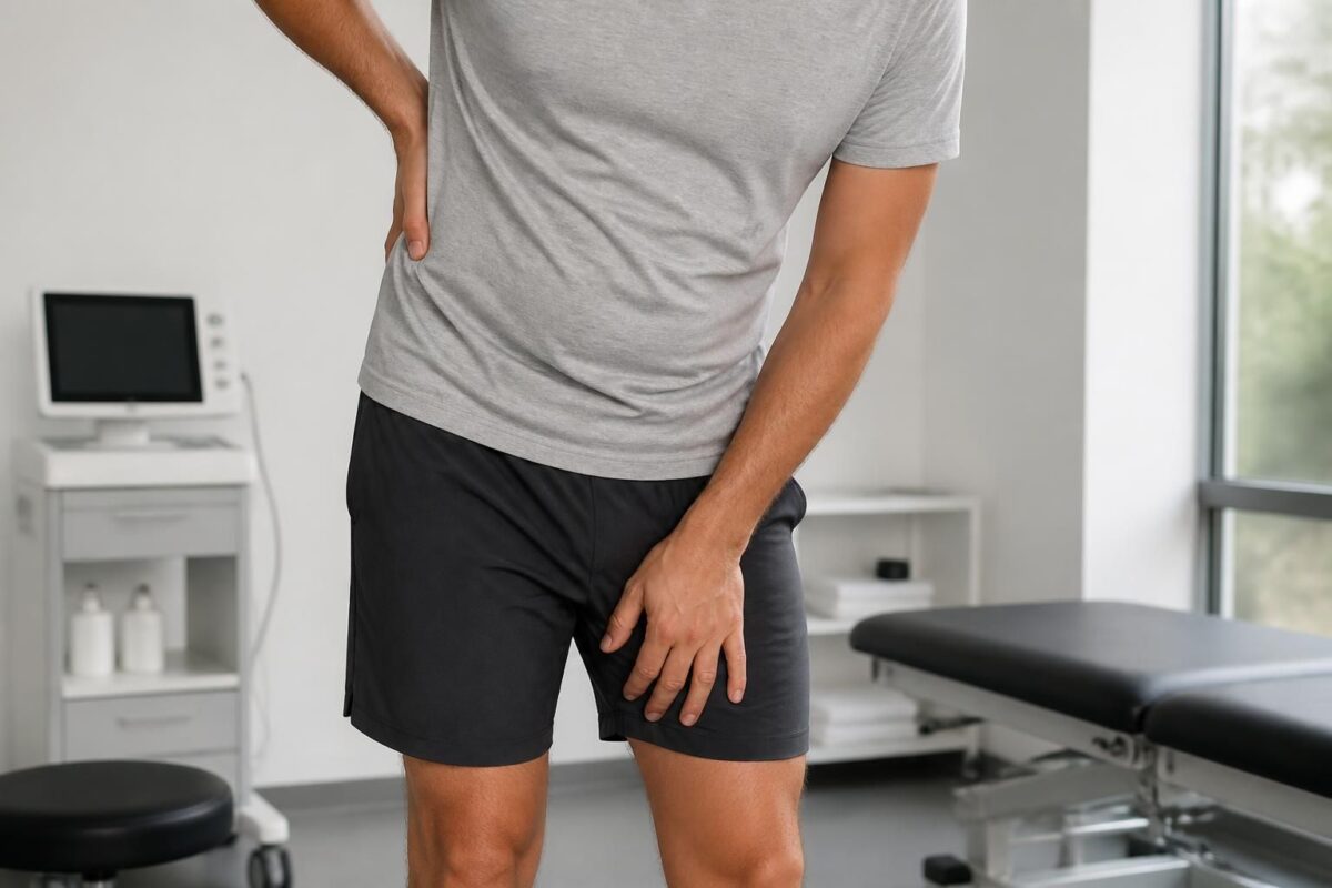
Sciatica and weakness in the thigh: possible lumbar cause
IN BRIEF Sciatica with thigh weakness is a possible manifestation of a lumbar cause irritating a nerve root (L4-L5, L5-S1). Advantages: a quick diagnosis and a targeted treatment plan with rehabilitation to regain strength and mobility and prevent recurrences. Operation:…
Practical support strategies
Adopting a progressive and multimodal approach allows you to quickly regain comfort and avoid relapses. The initial protocol combines Target rest, icing, and Stretching of the plantar fascia and calves To reduce inflammation and soften tissues. The use of Plantar orthoses Or suitable shoes can redistribute pressure and stabilize the arch of the foot, while helpers like gel heel pads protect the heel when walking. Simple exercises of strengthening and of Foot mobilization complete the program to improve stability and prevent recurrences. In some cases, targeted manual therapies and functional rehabilitation accelerate recovery.
When to consult and which signals to monitor
See quickly if the pain is Intense and disabling, if there is a marked swelling With redness and local heat, or if neurological signs appear (numbness, tingling, significant weakness). Pain that persists for more than two weeks despite the initial measures deserves a medical assessment. A general practitioner, podiatrist, or rheumatologist can determine whether a root pathology, tendinopathy or other heel condition is involved and refer to appropriate treatments, including advanced options when needed.
| Criterion | Quick Interpretation |
|---|---|
| Location and irradiation | S1 probable when the pain follows the posterior path of the calf to the arch and the heel; Other causes : Strictly local pain, under the heel without irradiation. |
| Beginning and timeline | plantar fasciitis : morning pain that improves with walking; S1 : fluctuating pain, without typical morning pattern. |
| Pain on waking | plantar fasciitis : marked morning pain; S1 : less specific when waking up. |
| Pain on palpation | localized pain at the insertion of the fascia under the heel; S1 : less targeted palpation; preponderant neurological signs. |
| Neurological signs | S1 : Weakness of the tip lifter and diminished achilles reflex; paresthesias; Fasciitis: No major neurological signs. |
| impact on walking | Fasciitis: Stronger pain at the beginning, then improvement; S1 : Pain that worsens on walking and exertion. |
| Useful clinical tests | SLR positive → radiculopathy; palpation fascia → fasciitis; Strength assessments and useful reflexes. |
| Action plan and when to consult | Positive SLR or neurological signs: specialist orientation; Otherwise conservative treatment (rest, ice, stretching, soles). Persistent pain >2 weeks: Consult. |
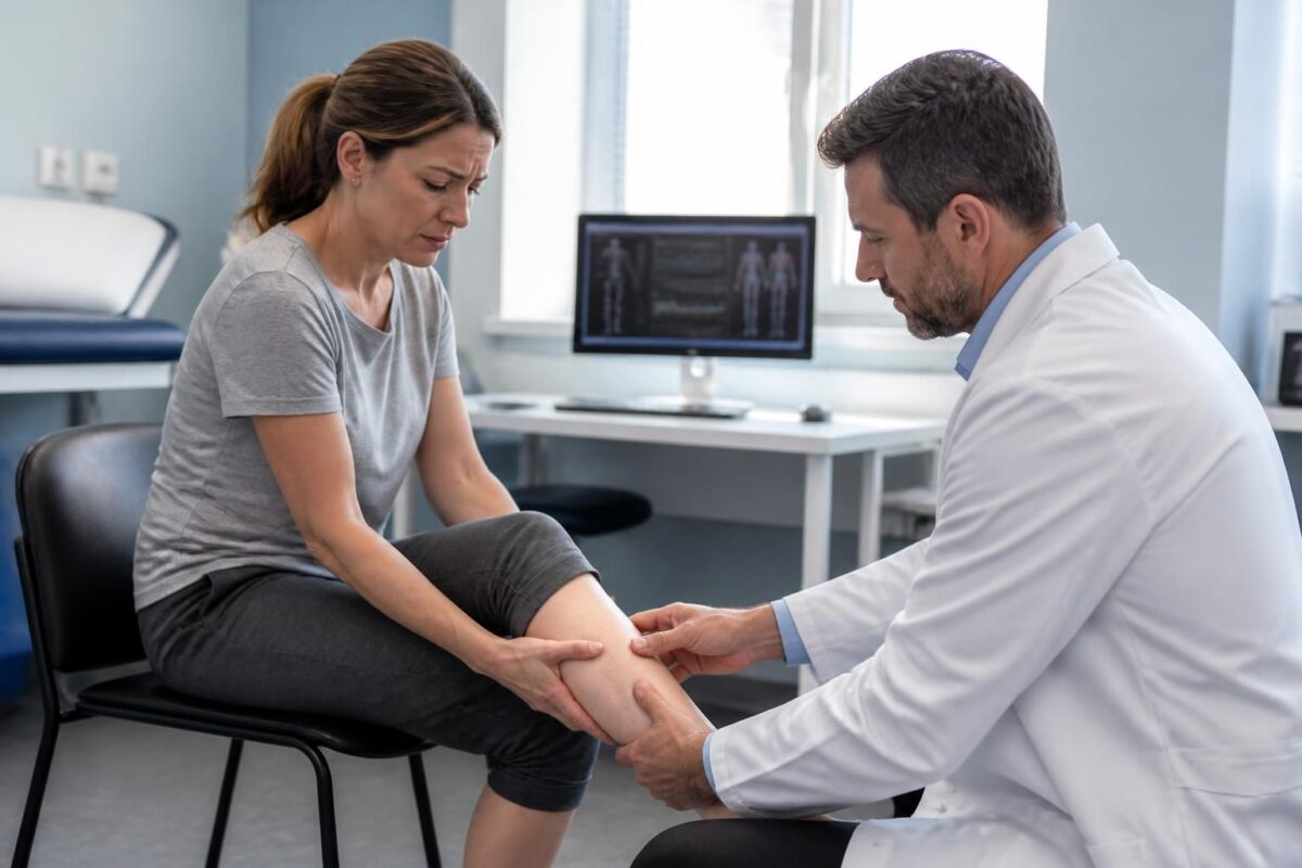
Sciatica and calf weakness: what should be monitored?
IN SHORT In short, sciatica and weakness in the calf describe a lumbar radiculopathy where pain follows the path of the sciatic nerve and may reach the calf. Benefits: a targeted non-surgical management, quick relief, and an improvement in mobility…

Sciatica and difficulty walking on tiptoes: possible S1 root
IN BRIEF In brief, sciatica with difficulty walking on tiptoes may reflect an involvement of the S1 lumbar root. What is it? a radicular pain that can limit foot elevation and walking on tiptoes. What are the benefits? an accurate…
Testimonials: Heel pain — S1 or other cause?
“My name is Claire, I am 39 years old and pain in the right heel that appeared after a period of overwork. The doctor mentioned a plantar fasciitis following repeated standing work and increased sporting activity. The program was simple and effective: targeted rest, ice, fascia and calves, and footwear offering good support. Today, the awakenings are no longer long and the pain moves away as I move. »
“I am Jean, 52 years old, and I felt a very localized pain in the center of the heel, especially on hard ground. A calcaneal thorn was considered after the initial examinations and the pain subsided thanks to adapted pads, foot orthoses and, if necessary, stimulation sessions, with a notable improvement over the weeks. »
“My name is Laura, I’m 29 and the pain is behind the heel after each running session. The diagnosis instead pointed to a Achilles tendinitis, related to overload. The treatment was based on progressive stretching of the calves, heel pads to reduce tension, and appropriate resumption of activity. Result: the pain has decreased sharply and I can resume my training gently. »
“My name is Pierre, 64, and I noticed pain in the back of the heel associated with a slight swelling and a feeling of warmth after a long walk. It was a Calcanean bursitis caused by friction and unsuitable shoes. The treatment consisted of removing irritating shoes, applying ice and using suitable protections; The pain decreased in one to two weeks. »
“My experience is that of Antoine, 46, with heel pain sometimes radiating in the foot after lower back pain. A S1 radiculopathy was considered when the symptoms of the back were added pain in the heel and lower back. Additional examinations made it possible to guide the treatment without resorting to surgery and to obtain progressive relief. »
“Finally, I’m Sophie, 50, and I found that the pain affects more right heel, probably by dominance and by increased mechanical load when moving. A biomechanical assessment has highlighted imbalances; Orthoses and postural advice were offered, which allowed for a gradual reduction in tensions and lasting improvement. »
Dr. Sylvain Desforges, a recognized osteopath and multidisciplinary practitioner, leads with Vision the Tagmed clinics and is actively involved in ACMA. His career is deeply rooted in innovation in health care and in the fight against Chronic pain. Expert in osteopathy, naturopathy And manual medicine, he favors an integrative approach that combines tissue analysis, the biomechanics of muscle chains and the reasoned use of modern technologies. Its mission is to provide care evidence-based to optimize the health and well-being of its patients.
In the context of heel pain, he applies himself to dissecting the mechanisms and possible origins, because a talalgia may come from local foot-level sources or reflect lumbar involvement, including Root S1. His approach aims to distinguish the causes affecting bone, muscular and nervous tissue, in order to offer a clear and personalized care course.
The most common causes he examines include the plantar fasciitis, thecalcaneal thorn, the Achilles tendinopathy and calcanean bursitis. Each has specific symptomatic profiles and requires appropriate treatment strategies. Dr. Desforges takes care not to neglect other less frequent but significant sources, such as fatigue fractures or systematic inflammatory conditions likely to impact the heel and foot.
when considering the involvement of S1, he advocates a rigorous clinical evaluation: pain radiating towards the lower limb, weakness or numbness in the territory of the sciatic nerve, pain aggravated on exertion and, sometimes, alteration of the Achilles reflex. This constellation may indicate lumbar radiculopathy, requiring a precise diagnostic approach and coordination with the possibilities of rehabilitation and neuroosteopathy. On the other hand, a typical plantar fasciitis is manifested in the morning by acute pain under the heel which fades a little after the first steps, before reappearing at the end of the day.
For Dr. Desforges, the differential diagnosis is a key step, because the proper identification of the origin makes it possible to avoid inappropriate treatments and to optimize the rehabilitation. Its practice is based on careful observation, targeted functional tests and, if necessary, an orientation towards additional examinations. He first favors conservative approaches adapted to the patient, while remaining attentive to any sign that could justify a quick specialized consultation.
In terms of care, it combines manual techniques and rehabilitation programs dedicated to the stability and recovery of the locomotor gesture. Advice on plantar orthosis, stretching and strengthening exercises, and appropriate management of overweight or reference overload factors are at the heart of its program. Moreover, Integration of advanced technologies such as spinal decompression, laser And shockwave therapies complete the approach when the pain persists or when the tissues requested require precise stimulation of tissue repair. This combination aims to reduce pain, restore function and prevent relapses, while remaining faithful to a process obvious and personalized.
Dr. Desforges will thus endeavor to clearly explain the potential causes of heel pain, the available treatment options and the objectives to be achieved to quickly regain fluid and pain-free mobility. Its approach is both therapeutic and educational, so that each patient understands the mechanisms at play and actively participates in their recovery.
This sheet explores the mechanisms of heel pain and distinguishes local causes from possible pain related to a S1 radiculopathy. It offers clinical benchmarks, management options and useful resources to quickly guide diagnosis and treatment.
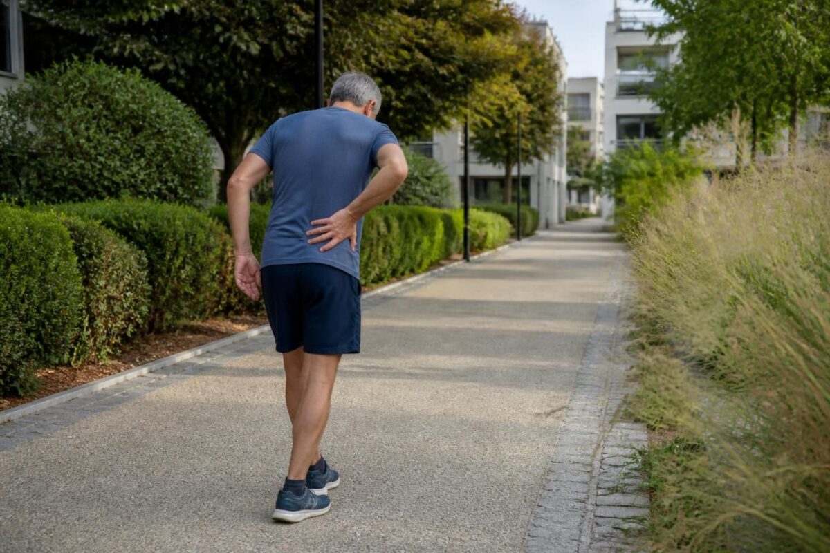
Sciatica and difficulty walking on heels: possible L5 root
IN BRIEF The sciatica associated with a difficulty walking on the heels is generally due to an involvement of the L5 root of the sciatic nerve, with a pathway that starts from the lower back, crosses the buttock, and goes…

Sciatica and difficulty lifting the foot: emergency or quick consultation?
IN BRIEF The drop foot is a weakness of the foot dorsiflexors that causes a steppage gait and may reveal sciatica with compression of the nerve in the lumbar region. When it is associated with sciatic pain and loss of…
Summary and key points
The sore heel may be due to a purely local problem, such as plantar fasciitis or thecalcaneal thorn, or root root associated with Root S1. The clinical indices that guide the examination include the pain distribution (does it ascend along the path of the nerve), the presence of Neurological signs (such as a reduction in the Achilles or paresthesia) and the functional context (overload, sports activity, posture). To deepen the relationship between sciatica and heel pain, consult the resources on the Sciatica and low back pain and the associated causes.
Among the most frequent local causes, we find the plantar fasciitis, often triggered by overload or repeated tension and characterized by acute pain on waking which improves temporarily and then returns at the end of the day. thecalcaneal thorn is a bone protrusion that can irritate adjacent tissues and cause localized pain in the center of the heel, especially on hard ground. The Achilles tendinitis and the Calcanean bursitis Complete the picture by bringing pain, swelling and sensitivity to the back of the heel, often in sports profiles or in case of unsuitable shoes. For additional perspectives and explanations, see Ameli Resources and other available guides on the subject.
In the presence of pain S1, the evaluation should aim to detect specific neurological signs and pain according to a root distribution (including buttocks and posterior part of the leg). Elements such as lingering pain despite rest, motor weakness or reflex change can direct towards radiculopathy and require a specialized approach (imaging, consultation in rheumatology or sports medicine). To cross information and mechanisms, consult dedicated resources such as those on the Causes of sciatica, the Piriform Syndrome, and the sheets dedicated to the Pain behind the thigh.
Support is based on simple and effective bases: Target rest, application of ice cream, stretches calves and plantar fascia, and choice of suitable shoes and orthoses. In case of persistence, options such as Plantar orthoses (tailor-made or prefabricated) and specific therapies can be considered, sometimes associated with techniques such as shockwave therapy for recalcitrant fasciitis. For further consultation and processing methods, please consult the AMELI resources and the specialized guides listed below.
Additional resources allow you to expand understanding of possible mechanisms and treatments: Ameli – Consultation and treatment, specialized articles on Heel pain, and practical sheets on the manifestations of Talalgia available on women’s diary. To explore the links between sciatica and associated pathologies, also find the resources dedicated to the Foraminal and sciatic stenosis.
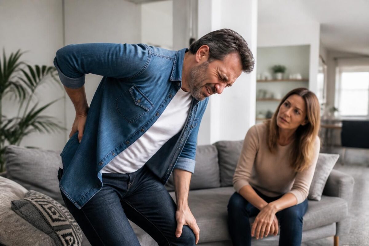
Sciatica and loss of strength: a sign not to be taken lightly
IN SHORT In short, sciatica is pain that follows the path of the sciatic nerve. A loss of strength in the leg is a sign that should not be taken lightly. It’s a warning indicating nerve damage that may require…

Sciatica and numb foot sensation: when to consult?
IN BRIEF In brief, sciatica and numbness in the foot refer to pain that follows the path of the sciatic nerve and may be accompanied by tingling or numbness in the foot. This summary specifies when to seek consultation, distinguishing…

Dr Sylvain Desforges, B.Sc., D.O., N.D., osteopath, is the founding president of TAGMED and of the ACMA association. He dedicates his practice to chronic spinal pain, low back pain and symptoms compatible with sciatica, particularly when pain radiates toward the buttock, leg, calf, foot or toes.
Through SOS Sciatique, he publishes educational content on sciatica symptoms, possible causes such as disc herniation, disc narrowing, foraminal stenosis or irritation of a lumbar nerve root, as well as non-surgical approaches that may be considered according to the evaluation. At Clinique TAGMED, care may include, depending on the case, motorized neurovertebral decompression, specific osteopathic care or precision percussion.
For an evaluation related to sciatica treatment in Montreal / Mount Royal or sciatica treatment in Terrebonne, please consult the services available at Clinique TAGMED.
Medical disclaimer
The information published on SOS Sciatique is provided for educational purposes only and does not replace a consultation, diagnosis, opinion or treatment provided by a physician or another authorized healthcare professional. The author of this article is neither a medical doctor nor a specialist in a medical specialty recognized by the Collège des médecins du Québec. The osteopathic, manual medicine or functional support approaches described on this site do not constitute medical acts and are not intended to replace medical follow-up.
Consult a physician or emergency service in cases of loss of bladder or bowel control, anesthesia in the saddle area, significant or progressive weakness in the leg or foot, fever, recent trauma, severe pain or rapid worsening of symptoms. For more details, please read our Legal Notice.