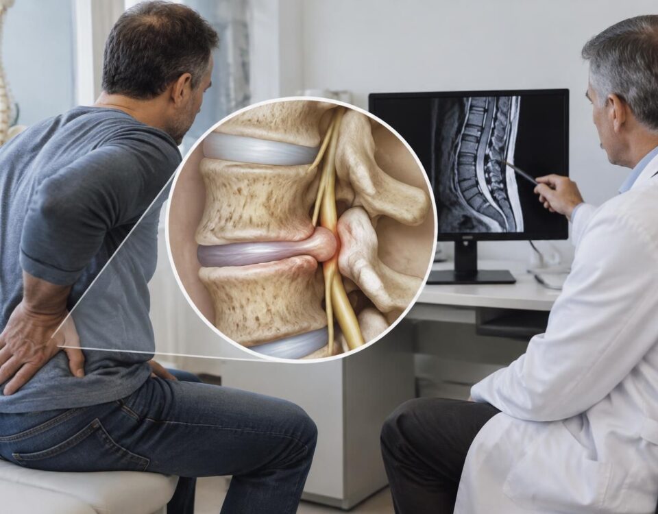



Sciatica and bar pain in the lower back with radiation
13/08/2026
Sciatica and long-distance driving: signs to watch for
13/08/2026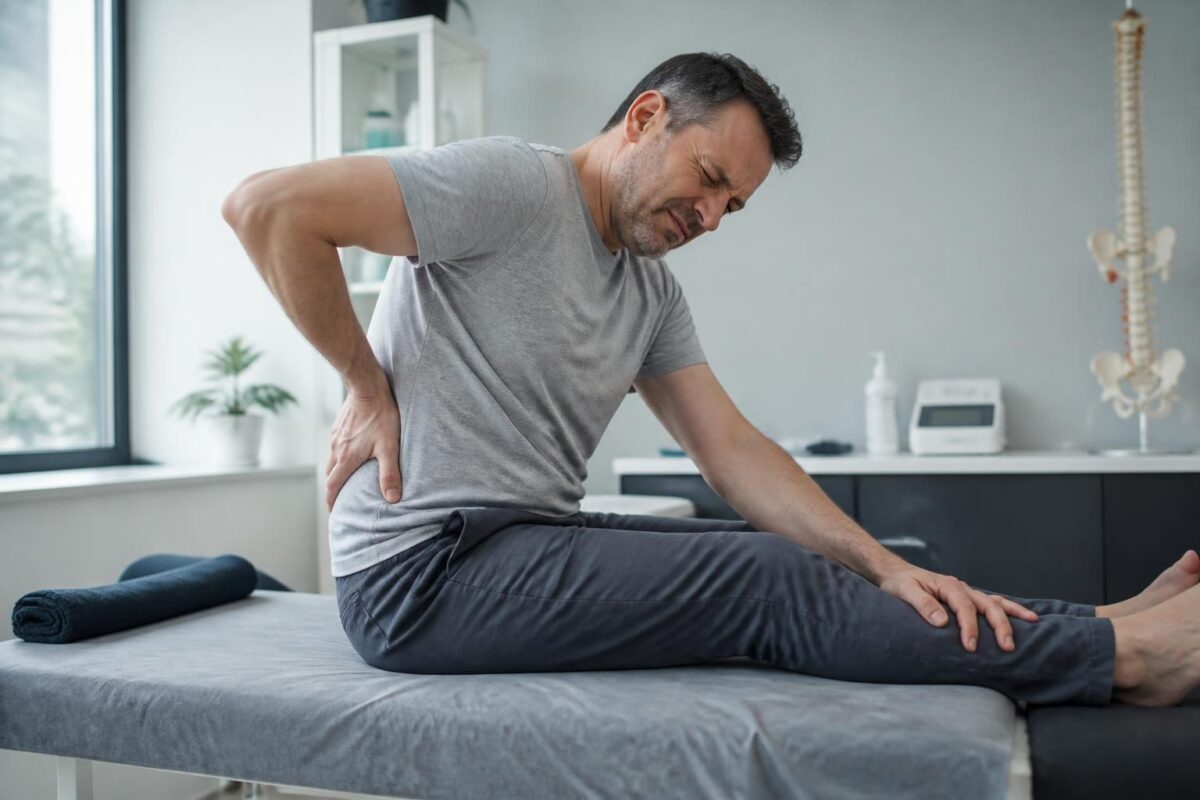
IN BRIEF
In brief, the extruded disc herniation is the protrusion of the nucleus pulposus through the ruptured annulus fibrosus, which can project into the spinal canal or the foramen and irritate or compress a nerve root, causing sciatica with pain radiating along the path of the sciatic nerve. This condition is distinguished from other causes of lower back pain by its radicular mechanism and its potential to lead to weakness or sensory disturbances. The main benefits of understanding this entity are to quickly guide management: effective conservative treatment (analgesics, anti-inflammatories, physiotherapy, injections) when there are no signs of urgency, and surgical indications in case of persistence or major neurological signs. The key mechanism is the compression or inflammation of the nerve root, with neurogenic pain that may worsen with effort or coughing. In Montreal, our clinics offer rapid assessments and tailored options for quicker relief and targeted rehabilitation. |
Extruded disc herniation and sciatica: clinical significance
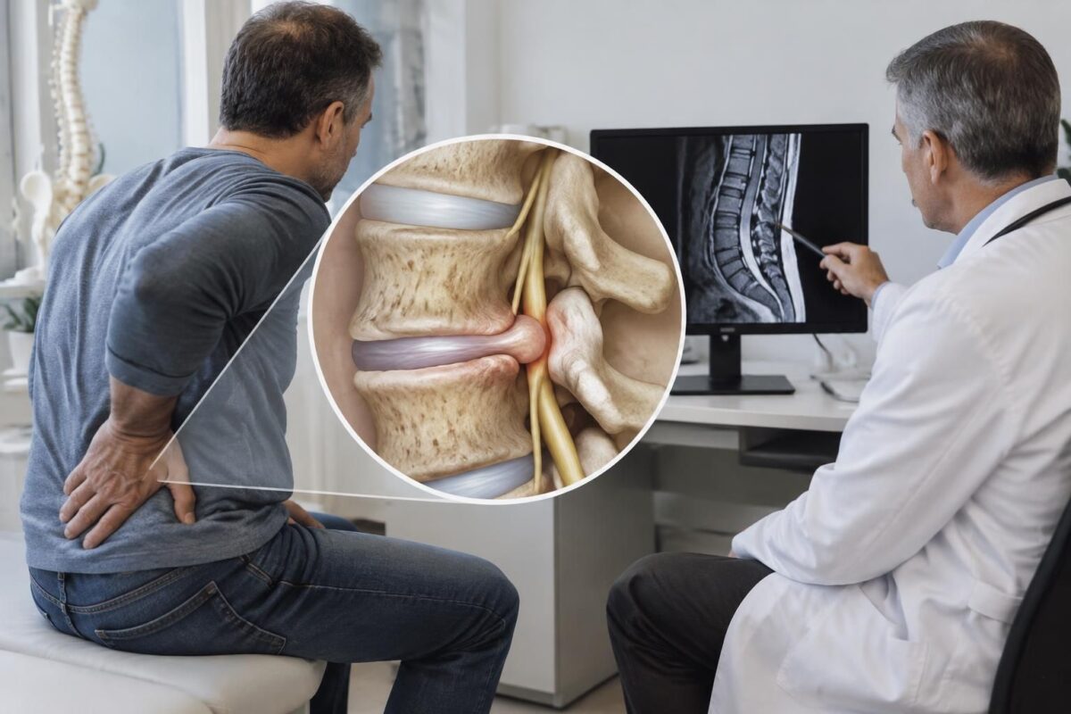
The extruded disc herniation corresponds to a more significant rupture of the fibrous ring with a fragment of the nucleus pulposus that can migrate and compress a nerve root, most often in the spinal canal. This form is frequently associated with radiculalgia causing neurogenic pain radiating along the path of the sciatic nerve and may be more symptomatic than contained forms.
Key signs and symptoms: unilateral lower back or buttock pain radiating to the leg and foot, burning or electric character, worsening with coughing or effort, and sometimes sensory disturbances or muscle weakness with decreased reflexes. Commonly localized at L5-S1, but can affect other levels depending on the nerve root affected.
Clinical management: the diagnosis relies on clinical evaluation and MRI imaging that confirms the extrusion and assesses nerve root compression. The initial treatment is conservative (analgesics/anti-inflammatories, physiotherapy focused on strengthening and neurodynamics, adapted activity). Epidural injections or adjunct therapies may be proposed if necessary. A surgical option (discectomy) is considered in cases of symptom persistence after 6 to 8 weeks, or in the presence of progressive motor deficit or cauda equina syndrome.
- Key signs: sudden radicular pain, lumbar radiation → leg, unilateral, sometimes sensory/motor deficit.
- Imaging: MRI is essential to confirm the extrusion and assess compression.
- Treatment pathway: initial medical treatment + rehabilitation; surgery if failure or neurological emergency.
This article, written by an expert in osteopathy and manual medicine, clarifies the clinical significance of the extruded disc herniation associated with sciatica, describes the mechanisms involved, diagnostic criteria, and management options, while guiding the care pathway towards tailored solutions at TAGMED clinic.
Definition and mechanisms
An extruded disc herniation occurs when the nucleus pulposus of an intervertebral disc breaches a fissure in the fibrous ring and is located in the spinal canal or in the foramen, forming a free disc fragment. This situation is distinguished from simple protrusion by the fact that the disc material has crossed the capsule and can migrate. The extrusion can irritate or compress a nerve root, causing radicular pain. Sciatica most often results from this compression or associated inflammation of the L5 or S1 root, with a painful path following the sciatic nerve to the lower limb.
Contributing factors include disc degeneration, mechanical stress, and the inflammatory process. An extrusion can be central or lateral, and the fragment can move within the spinal canal or towards the foramina, which influences the clinical picture and therapeutic decisions.
Clinical signs and diagnosis
Sciatica associated with a disc extrusion is typically unilateral and manifests as intense, burning, or electric radicular pain that travels along the path of the affected nerve and may involve the buttock, thigh, leg, and sometimes the foot. The pain is often exacerbated by coughing, exertion, or defecation. Neurological signs such as hypoesthesia, motor weakness, or altered reflexes may be present when the root is severely compressed.
The clinical examination looks for signs of Lasègue and assesses motor and sensory deficits. Imaging is a key component of the diagnosis: MRI is the reference exam to confirm an extrusion and specify the location of the fragment (e.g., L4-L5, L5-S1). CT can screen for particular cases or complement the assessment when MRI is inconclusive. In some contexts, the combination of imaging and clinical information allows differentiation between an extrusion and a protrusion and clarifies the risk of serious complications.
Imaging and differential diagnosis
Imaging plays a central role in distinguishing disc extrusion from other forms of herniation and spinal pathologies. An extrusion may coexist with foraminal stenosis or signs of periradicular inflammation. The differential diagnosis includes other causes of radicular low back pain and vascular or tumor pathologies that may mimic radiculopathy. For patients presenting with progressive symptoms or significant neurological signs, rapid assessment is recommended to prevent complications, such as cauda equina syndrome.
To explore approaches based on your symptoms and context, consult specialized resources on sciatica and disc extrusions:
- Sciatica in Terrebonne — how to choose a safe approach according to your symptoms
- Central disc herniation and sciatic pain — what you need to understand
- Burning pain in the leg — is it sciatica?
- Sciatica in Montreal — pain in the foot, numbness, and tingling
- Sciatica in Terrebonne — safe approach according to your symptoms
Management and therapeutic options
The management of an extruded disc herniation with sciatica relies on a multimodal approach tailored to the patient. Treatment usually begins with symptomatic measures and careful monitoring. Possible options include medication treatments (analgesics and anti-inflammatories), targeted injections, and non-surgical interventions when appropriate. The neurovertebral decompression and specific osteopathic techniques may be part of the overall strategy, under specialized supervision and within a multidisciplinary team.
In cases where symptoms persist despite appropriate management, or in the presence of progressive motor deficits or signs suggesting emergency, surgical intervention may be considered. The surgery aims to remove the extruded fragment and relieve the compression, with rapid improvement rates observed in some patients and a gradual recovery in the long term.
Comparative table of therapeutic approaches
| Treatment | Relative efficacy index | Remarks |
|---|---|---|
| Anti-inflammatories and analgesics (NSAIDs, paracetamol) | Initially high | Used as a first resort; effects vary depending on the patients. |
| Epidural infiltrations | Moderate to high | Can quickly reduce radicular pain; individual assessment needed. |
| Motorized neurovertebral decompression (at TAGMED clinic) | Good impressive for some cases | Non-surgical option when suitable; integrated into overall management. |
| Physical therapy | Variable effectiveness | To be considered as a complement if beneficial; prioritize targeted therapies and clinical options. |
| Surgical discectomy | Rapid and significant in some patients | Reserved for refractory patients or those with clear surgical indications; postoperative rehabilitation necessary. |
To discuss options specific to your case and access specialized services, contact the TAGMED Clinic in Montreal or Terrebonne. To learn more about the proposed approaches, visit the following resources:
- TAGMED Clinic
- Neurovertebral decompression
- Disc herniation — associated resources
- Extruded disc herniation — information
- SOS Sciatica and associated resources
Key points for clinical practice
A sciatica related to an extrusion may be present even without a visible disc herniation on imaging and vice versa. In-depth clinical evaluation remains central, with particular attention paid to signs of severity and “red flags” requiring rapid management. Imaging is useful when clinical evolution is slow or when neurological symptoms appear or worsen. A multidisciplinary approach, combining physician, osteopath, and, if necessary, a surgeon, is often the key to effective and tailored management.
Dr. Sylvain Desforges, B.Sc., D.O., N.D., osteopath, is the founding president of Through SOS Sciatica, he publishes educational content about the symptoms of sciatica, possible causes such as disc herniation, disc pinching, foraminal stenosis, or irritation of a lumbar nerve root, as well as non-surgical approaches that may be considered based on the assessment. At TAGMED Clinic, care may include, depending on the case, motorized neurovertebral decompression, specific osteopathy, or a precision impactor. For an evaluation related to Medical disclaimer The information published on SOS Sciatica is provided for educational purposes and does not substitute for a consultation, diagnosis, advice, or treatment performed by a physician or other licensed health professional. The author of this article is neither a doctor nor a specialist in a medical field recognized by the Collège des médecins du Québec. The approaches of osteopathy, manual medicine, or functional support described on this site do not constitute medical acts and are not intended to replace medical follow-up. Consult a doctor or an emergency service in case of urinary or bowel incontinence, anesthesia in the saddle region, significant or progressive weakness of the leg or foot, fever, recent trauma, severe pain, or rapid deterioration. For more details, please read our
About the author
TAGMED
and of the ACMA association. He dedicates his practice to chronic spinal pain, low back pain, and pain compatible with sciatica, particularly when the pain radiates to the buttock, leg, calf, foot, or toes.
sciatica treatment in Montreal / Mont-Royal
or to
sciatica treatment in Terrebonne,
consult the available services at the
TAGMED Clinic.
Legal notice.
Dr. Sylvain Desforges, Osteopath, thanks you for your trust and invites you to consult the resources and care offered by TAGMED for an evaluation tailored to your case.
Medical disclaimer: The information and advice provided on this site do not replace the opinion, diagnosis, or treatment of a healthcare professional. Please note that Dr. Sylvain Desforges, osteopath, is neither a medical doctor nor trained in a medical specialty as defined by the Collège des médecins du Québec. Manual medicine, functional medicine, and sports medicine as described on this site exclude any treatment or medical diagnosis made by a physician or medical specialist. Always consult your physician for any medical questions. For more details, please read our complete Legal Notice.
- Extruded disc herniation: extrusion of the nucleus pulposus through the annulus; protrusion into the spinal canal or foramina.
- Clinical signs: marked radicular pain; possible motor or sensory deficit.
- Common locations: L4-L5 or L5-S1.
- Diagnosis: MRI to confirm; clinical correlation essential.
- Management: NSAIDs and analgesics, physiotherapy; surgery considered if motor deficit or persistence >6–8 weeks.
- Sciatica: neuralgic pain radiating to the buttock and leg, following the path of the sciatic nerve.
- Characteristics: unilateral, burning pain; aggravated by effort, coughing, and defecation.
- Associated signs: hypoesthesia, weakness, diminished reflexes; positive Lasègue sign possible.
- Diagnosis: clinical examination; imaging if persistence or suspicion of red flags.
- Management: NSAIDs and analgesics; physiotherapy and injections; surgery if disabling sciatica or refractory to treatment.

This text presents the essential clinical issues of extruded disc herniation and sciatica, distinguishing the mechanisms, signs, and management options. It aims to clarify the diagnosis and guide a patient-centered therapeutic strategy focused on functional effectiveness.
Definition and clinical significance
An extruded disc herniation occurs when the nucleus pulposus of the intervertebral disc breaches the annulus fibrosus and projects outside its confinement while remaining connected to the disc or forming a free fragment in the spinal canal. This situation can exert pressure or irritation on adjacent nerve roots, particularly in the lumbar regions, and is often associated with pain radiating along the path of the affected nerve. In practice, it differs from disc protrusion, where the material remains largely within the same level, and sequestrated fragment, when the fragment has completely separated. The clinical severity depends on the location of the fragment and the degree of radicular conflict.
Clinical signs and differential diagnosis
Characteristic signs
The main complaint is radicular pain that follows the path of the compressed nerve, often with radiation to the buttock, leg, and sometimes the foot. This pain is typically neurogenic in nature and can be aggravated by coughing, sneezing, or effort. Motor or sensory symptoms, such as hypoesthesia, muscle weakness, or diminished reflexes, may accompany the radicular conflict and direct towards a confirmed nerve injury upon examination.
When imaging is useful
The use of magnetic resonance imaging is indicated when the clinical picture is persistent, accompanied by marked neurological signs, or there is doubt about a complication. However, it should be kept in mind that imaging can reveal a disc herniation without symptoms, and the absence of deficit does not rule out radiculopathy if the clinical picture suggests it.
Management: practical approach
Initial non-surgical approach
Management typically begins with pain control and functional improvement, using anti-inflammatories, appropriate analgesics, and physiotherapy focused on neurodynamics, trunk strengthening, and stretching exercises as tolerated. Postural education and maintaining suitable physical activity play a key role in preventing recurrences and in the overall improvement of spinal stability.
Surgical indications and follow-up
Surgery may be considered in cases of treatment-resistant sciatica after a period of observation or in the presence of progressive motor deficit, signs of severe compression, or suspected cauda equina. The surgery aims to remove the exposed fragment and relieve the nerve root while preserving healthy structures as much as possible. The use of minimally invasive techniques can facilitate recovery and limit hospital stay, while generally allowing a return to activities within a few weeks to a few months.
Prognosis and factors influencing recovery
Evolution and expectations
Disc extrusions often have potential for spontaneous resorption and symptom improvement over time, but the duration and intensity vary according to the patient and the location of the fragment. Prognosis is influenced by the severity of radicular conflict, age, fitness level, and promptness of management. A multidisciplinary strategy involving a physician, physiotherapist, and, if necessary, surgeon maximizes the chances of functional recovery and pain reduction.
Prevention and maintenance of spinal health
Promoting trunk strengthening, maintaining a healthy weight, adopting appropriate lifting techniques, and preserving suitable mobility help reduce stress on the discs and limit painful episodes. Targeted and progressive rehabilitation contributes to preventing recurrences and supporting functional quality of life.
| Aspect | Clinical significance |
|---|---|
| Definition | Extruded disc herniation: protrusion of the nucleus pulposus through the annulus that may compress a root; Sciatica: radicular pain following the path of the involved nerve. |
| Location and course | Extruded disc herniation typically located at L4-L5 or L5-S1 with low back pain and possible irradiation; Sciatica: radiating pain along the path of the sciatic nerve down to the foot. |
| Mechanism | Extruded disc herniation: mechanical compression and radicular inflammation; Sciatica: radiating neuropathic pain related to radicular involvement. |
| Neurological signs | Extruded disc herniation: possible neurological deficit, infrequent; Sciatica: hypoaesthesia, weakness, or decreased reflexes when the root is affected. |
| Imaging | Extruded disc herniation: MRI or CT helpful to confirm the herniation and its relationship; Sciatica: may exist even without imaging showing a herniation. |
| Diagnosis and examination | Extruded disc herniation: targeted clinical examination and imaging if persistent or signs of alert; Sciatica: Lasègue and motor/sensory assessment guide the diagnosis. |
| Initial management | Extruded disc herniation: relative rest, analgesics, and rehabilitation; Sciatica: medication treatment and physiotherapy tailored to severity. |
| Surgical indications / emergency | Surgery considered after treatment failure or motor deficit for extruded disc herniation; Sciatica may require discectomy when severe pain or persistent deficit occurs. |
Testimonials on Extruded Disc Herniation and Sciatica: Clinical Significance
“I had a shooting pain that started from the buttock and radiated down to the foot. The doctor diagnosed me with extruded disc herniation with compression of a nerve root and triggering of sciatica. The clinical significance was clear: the pain follows the path of the nerve and may be accompanied by tingling or weakness. This understanding helped me accept the care plan and stay active despite the symptoms.”
“The MRI confirmed that the episode was related to a left extruded disc herniation and sciatica. The clinician explained that even though the pain comes from the nerve, the goal of treatment is to reduce the inflammation around the root and support the back through exercises. I learned that distinguishing between mechanical low back pain and radicular pain is crucial for choosing non-surgical approaches and avoiding unnecessary treatments.”
“I thought that all sciatica came from a herniation, but the doctor clarified that radicular pain can exist without a correlated image and that other mechanisms may be at play. This clinical significance helped me not to panic when the MRI was inconclusive and to pursue a rehabilitation plan focused on the nerve and trunk.”
“After a few weeks, the physiotherapy work and decompression techniques improved my mobility. I was explained that, in some cases, an extruded disc herniation can benefit from a conservative approach and that surgery is only considered if symptoms persist or worsen. Understanding the clinical significance made decisions clearer and less anxiety-inducing.”
“I realized that sciatica can be intermittent and that factors like posture and movement influence the intensity. The doctor stressed that clinical assessment is essential and that imaging is a tool among others to guide treatment. This approach helped me adopt postural habits and exercises that protect the back in the long term.”
Clinical Significance of Extruded Disc Herniation and Sciatica
Dr. Sylvain Desforges is a recognized osteopath, expert in naturopathy and manual medicine, who serves as the founder president of TAGMED clinics and the ACMA association. His career highlights a focus on innovation in healthcare and a resolutely patient-centered approach to understanding and managing chronic pain. His work is based on thorough patient assessment, therapeutic education, and the thoughtful use of advanced technologies to improve quality of life, especially for those suffering from low back and radicular pain.
Clinically, he precisely distinguishes between extruded disc herniation and manifestations of sciatica, reminding that each phenomenon is based on specific mechanisms. The extruded herniation is defined by the migration of the nucleus pulposus beyond the annulus, sometimes with radicular compression and local inflammatory reaction. Sciatica is a neuritic pain that follows the path of the sciatic nerve, most often related to irritation or compression of a nerve root (typically L5 or S1), and may exist even in the absence of marked herniation on imaging. This distinction guides the therapeutic strategy and the objectives for preventing recurrences.
In his approach, Dr. Desforges favors a patient-centered methodology, combining a detailed clinical evaluation (radicular pain tests, motor and sensory assessment, and identification of neurological alert signs) and targeted imaging when the picture persists or is accompanied by deficits. He emphasizes that many disc herniations can be asymptomatic and that sciatic pain can occur without marked radiological abnormality, reinforcing the importance of a dynamic diagnosis and case-by-case follow-up.
According to him, management is multidisciplinary and progressive. For simple low back pain, protocols emphasize trunk strengthening, enhancing mobility, and postural education, while avoiding prolonged rest. In cases of extruded herniation associated with significant radiculopathy, the therapeutic arsenal combines active advice, targeted manual therapies, and non-invasive interventions (physiotherapy, neurodynamic rehabilitation, infiltrations when necessary), with regular reassessment to determine the possibility of surgical intervention or adjustment of the treatment plan, prioritizing low-risk and effective solutions.
Furthermore, the integration of advanced technologies is an integral part of its approach. Motorized spinal decompression, laser, and shockwave therapies are used as levers to modulate pain, promote tissue recovery, and improve nervous and musculoskeletal function. This technological dimension is part of an approach based on evidence, with clinical protocols aimed at optimizing patient function and safety while respecting individual healing rhythms.
Through his activities at the helm of TAGMED clinics and ACMA, Dr. Desforges promotes care that combines knowledge, technique, and personalized support. His mission is to improve the health and well-being of individuals suffering from low back and radicular pain, by reconciling osteopathic approach, manual medicine, and modern technologies to facilitate a quick and sustainable return to activity, within a framework of continuous and tailored care.
This text provides a clear analysis of extruded disc herniation and sciatica, specifying their clinical significance, underlying mechanisms, and implications for management. It highlights the distinctions between protrusion, extrusion, and radiculopathy, and describes therapeutic options based on severity and treatment response.
Clinical significance of extruded disc herniation and sciatica
In the context of extruded disc herniation, the nucleus pulposus breaches the annulus fibrosus and can end up in the spinal canal or at the intervertebral foramina, with a potential risk of displacement and localized inflammation. This situation is often associated with sciatica when the nerve root is compressed or irritated, producing neurogenic pain that follows the path of the affected nerve down to the lower limb.
The clinical mechanism typically favors radiating pain, sometimes burning, which may be accompanied by paresthesia, weakness, or motor deficit depending on the level of the affected root. The pain is generally unilateral and may be exacerbated by coughing, straining, or defecation, reflecting the inflammatory and mechanical radicular component.
It is crucial to distinguish extruded disc herniation from simple protrusions and to understand that the correlation between imaging and symptoms can vary: an extrusion does not necessarily imply major neurological loss and some low back pains can persist even in the absence of obvious pathological imaging. For practical aspects and mechanisms, see specialized resources on disc herniation and low back pain.
Diagnosis and clinical assessment
The diagnosis relies on clinical examination, particularly the search for signs of radiculopathy and motor or sensory deficits, as well as reflex tests. Imaging is useful to confirm the presence and location of an extruded herniation, but does not replace clinical evaluation. MRI is the reference examination to visualize the extent of the extrusion and its relationship with the nerve roots; however, a disc herniation can be asymptomatic and radicular pain can exist without a clear corresponding image.
In practice, the therapeutic decision is based on the association between the clinical picture and imaging results, as well as on the temporal evolution of the symptoms. Online resources provide additional guidance on sciatica and non-surgical treatment alternatives, especially in complex or chronic cases.
Therapeutic implications and management
Initial management often prioritizes non-surgical approaches: anti-inflammatories and analgesics, physiotherapy for MOBILIZATION, strengthening the core, and neurodynamic exercises, as well as postural education measures and weight management. In some cases, epidural infiltrations or injections may be considered to reduce inflammation and radicular pain.
Surgery is generally reserved when symptoms persist beyond 6 to 8 weeks despite appropriate treatment, or in the presence of progressive motor deficits, cauda equina syndrome, or neuro-surgical emergencies. The intervention may include a discectomy or targeted decompression, and minimally invasive techniques with regional anesthesia allow for rapid recovery in most cases. Some resources also discuss non-surgical alternatives like neurovertebral decompression and associated options for sustainably alleviating chronic disc herniations.
It is useful to consult specialized resources to clarify choices between medical treatment, decompression, and surgery, and to understand developments in therapeutic approaches for chronic disc herniations and refractory sciatica.
Prevention, rehabilitation, and practical framework
Prevention relies on maintaining appropriate physical activity, strengthening the abdominal and lumbar belts, and having a correct posture when standing or sitting. Strengthening and stretching exercises should be progressive and adapted to pain: they aim to stabilize the spine and reduce tension on the discs and roots. Using an appropriate lifting technique, with hips and knees bent, helps to limit the risk of injury and painful episodes.
For those suffering from sciatica associated with extruded disc herniation, a multimodal and individualized approach remains key: quickly assessing signs of severity, establishing targeted rehabilitation, and considering complementary interventions based on clinical response and imaging results.
To deepen the understanding of the mechanisms, non-surgical options, and surgical indications, here are some useful additional resources: Herniated disc (Vidal), Sciatica – Doctissimo, Herniated disc – MSD Manual, Neuro-vertebral decompression and herniated discs – SOS Sciatica, Decompression without surgery – SOS Sciatica, Sciatica and leg pain, Link between herniated disc and sciatica.
For any questions regarding the evolution and management, it is recommended to rely on a multidisciplinary approach and personalized follow-up with health professionals, in order to optimize diagnosis and guide therapeutic decisions according to individual needs.
Disc protrusion and disc pinching: combined mechanisms
IN BRIEF A disc protrusion is a bulging of the disc, while a disc narrowing refers to a decrease in its height. These changes can coexist and sometimes irritate a nerve root, leading to pain or radiation, but they can…

Disc herniation and associated foraminal stenosis
IN BRIEF A disc protrusion associated with foraminal stenosis occurs when a bulging disc coexists with a narrowing of the opening through which a nerve root passes. This association can lead to lower back pain radiating into the leg, tingling,…

Spinal disc protrusion and nerve inflammation
IN BRIEF A disc protrusion is a bulging of the disc that can irritate or compress a nerve, without necessarily causing symptoms. When it leads to nerve inflammation, it can cause pain in the back or neck, sometimes accompanied by…

Disc protrusion and irritation of L5
IN SHORT A disc protrusion is a bulging of the disc that can irritate the L5 root and cause lower back pain radiating to the buttock, leg, or foot, sometimes with tingling or weakness. MRI alone is not sufficient to…

Disc protrusion and pain after twisting the back
IN BRIEF A disc protrusion is a bulging of the disc that can contribute to pain after a back twist, but it alone does not confirm the cause. The pain can remain in the lower back or radiate to the…

Disc herniation and pain after lifting
IN BRIEF A disc protrusion is a bulging of a disc between the vertebrae. Pain that appears after lifting a load does not prove that it is the cause: the MRI must be interpreted alongside the symptoms and the clinical…

Disc herniation and lumbar flexion: why caution is necessary
IN BRIEF A disc protrusion is a bulge of the intervertebral disc, which can be silent or irritate a nerve. Lumbar flexion is not necessarily dangerous but can worsen symptoms in some individuals. Adapting movements, avoiding those that aggravate pain,…

Disc herniation and pain that worsens in the car
IN BRIEF A disc protrusion is a bulging of a disc that can irritate a nerve. In a car, prolonged sitting and vibrations can exacerbate back, buttock, leg, or arm pain. Regular breaks, an adapted posture, and personalized management can…

Dr Sylvain Desforges, B.Sc., D.O., N.D., osteopath, is the founding president of TAGMED and of the ACMA association. He dedicates his practice to chronic spinal pain, low back pain and symptoms compatible with sciatica, particularly when pain radiates toward the buttock, leg, calf, foot or toes.
Through SOS Sciatique, he publishes educational content on sciatica symptoms, possible causes such as disc herniation, disc narrowing, foraminal stenosis or irritation of a lumbar nerve root, as well as non-surgical approaches that may be considered according to the evaluation. At Clinique TAGMED, care may include, depending on the case, motorized neurovertebral decompression, specific osteopathic care or precision percussion.
For an evaluation related to sciatica treatment in Montreal / Mount Royal or sciatica treatment in Terrebonne, please consult the services available at Clinique TAGMED.
Medical disclaimer
The information published on SOS Sciatique is provided for educational purposes only and does not replace a consultation, diagnosis, opinion or treatment provided by a physician or another authorized healthcare professional. The author of this article is neither a medical doctor nor a specialist in a medical specialty recognized by the Collège des médecins du Québec. The osteopathic, manual medicine or functional support approaches described on this site do not constitute medical acts and are not intended to replace medical follow-up.
Consult a physician or emergency service in cases of loss of bladder or bowel control, anesthesia in the saddle area, significant or progressive weakness in the leg or foot, fever, recent trauma, severe pain or rapid worsening of symptoms. For more details, please read our Legal Notice.