


Sciatica and pain that worsens when walking
23/10/2026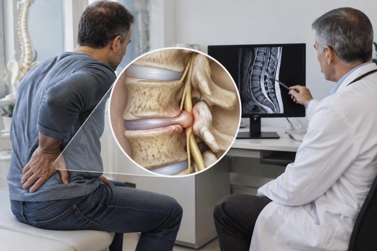
IN BRIEF
| A disc protrusion is a bulging of the disc, while a disc narrowing refers to a decrease in its height. These changes can coexist and sometimes irritate a nerve root, leading to pain or radiation, but they can also be asymptomatic. Understanding them helps in interpreting imaging alongside clinical examination and guiding appropriate management. |
A disc narrowing corresponds to a decrease in the height of the disc between two vertebrae, often related to its wear and loss of elasticity. A disc protrusion is a bulge of the disc outward. These two changes can coexist: a thinned disc can deform under stress without automatically meaning that a nerve is compressed.
Their significance depends on their location and their correlation with symptoms. Nerve irritation can notably cause pain that radiates into the buttock or leg, tingling, or numbness. Interpretation must take into account the clinical examination, not just the imaging. Progressive weakness, loss of bladder or bowel control, or anesthesia of the saddle area require urgent medical evaluation.
A disc protrusion is a bulging of the disc between two vertebrae. A disc narrowing refers to a decrease in the height of this space, often related to changes in the disc. Both observations can be present at the same time, but they do not always cause pain. Their significance depends on their location, their effect on nearby structures, and their correlation with symptoms.
What do disc protrusion and disc narrowing mean?
Disc protrusion corresponds to a bulge
Each intervertebral disc consists of a soft center called the nucleus pulposus, surrounded by a tough layer, the annulus fibrosus. A protrusion occurs when the contour of the disc overflows from its usual shape. The disc generally remains contained, even if its wall may be weakened.
The term “protrusion” describes a shape observed in imaging. It does not, on its own, indicate whether the disc irritates a nerve or explains a pain. To better understand the terms used in a report, consult our article on what the MRI can show in the presence of a disc herniation and sciatica.
Disc narrowing indicates a decrease in height
Disc narrowing corresponds to a reduction in height between two vertebrae. It may occur when the disc loses some of its hydration or its ability to distribute stress. The term describes a sign visible on an X-ray or other imaging. It does not necessarily indicate nerve compression.
The loss of height may also change the distribution of pressures on the joints and surrounding tissues. Its effect varies depending on the level of the spine, the extent of the change, and other clinical findings.

Disc herniation and associated foraminal stenosis
IN BRIEF A disc protrusion associated with foraminal stenosis occurs when a bulging disc coexists with a narrowing of the opening through which a nerve root passes. This association can lead to lower back pain radiating into the leg, tingling,…

Spinal disc protrusion and nerve inflammation
IN BRIEF A disc protrusion is a bulging of the disc that can irritate or compress a nerve, without necessarily causing symptoms. When it leads to nerve inflammation, it can cause pain in the back or neck, sometimes accompanied by…
How can these two mechanisms combine?
A disc that loses height can also change shape and bulge outward. A protrusion and a disc narrowing can therefore coexist. They may reflect different aspects of disc evolution, but one does not automatically prove that the other is the cause.
The narrowing can reduce the space available between the vertebrae. A protrusion can, depending on its direction and location, come closer to a nerve root. The combination may then contribute to nerve irritation or compression. However, imaging alone does not establish the link with certainty.
The proximity of a nerve matters more than just the report wording
A protrusion distant from the nerves can remain asymptomatic. Conversely, a smaller protrusion can become symptomatic if it is near an already irritated nerve root. Local inflammation can also contribute to pain, even if the visible compression seems moderate.
The affected level helps interpret the signs. In the lower back, nerve irritation can lead to pain that radiates into the buttock or leg. Some people experience tingling, numbness, or weakness. To learn more about the differences between lumbar levels, check the pages on sciatica related to L4-L5 and L5-S1 and on pain towards the top of the foot associated with L4-L5.

Disc protrusion and irritation of L5
IN SHORT A disc protrusion is a bulging of the disc that can irritate the L5 root and cause lower back pain radiating to the buttock, leg, or foot, sometimes with tingling or weakness. MRI alone is not sufficient to…

Disc protrusion and pain after twisting the back
IN BRIEF A disc protrusion is a bulging of the disc that can contribute to pain after a back twist, but it alone does not confirm the cause. The pain can remain in the lower back or radiate to the…
What symptoms can accompany these changes?
A protrusion or disc narrowing may be discovered without pain. When symptoms occur, they depend on the affected area and the structures involved. Pain may remain localized in the back or neck. It can also radiate to a buttock, leg, shoulder, or arm if a nerve root is irritated.
In the lumbar region, pain following the path of the sciatic nerve may be accompanied by tingling or altered sensitivity. However, the presence of a narrowing does not automatically mean it is sciatica. Consult our information on sciatica and lumbar disc herniation and on sciatica associated with disc narrowing.

Disc herniation and pain after lifting
IN BRIEF A disc protrusion is a bulging of a disc between the vertebrae. Pain that appears after lifting a load does not prove that it is the cause: the MRI must be interpreted alongside the symptoms and the clinical…

Disc herniation and lumbar flexion: why caution is necessary
IN BRIEF A disc protrusion is a bulge of the intervertebral disc, which can be silent or irritate a nerve. Lumbar flexion is not necessarily dangerous but can worsen symptoms in some individuals. Adapting movements, avoiding those that aggravate pain,…
Why do these changes occur?
Discs evolve over time. They may lose some of their hydration and become less flexible. Repeated stresses, certain efforts, prolonged postures, and trauma can also contribute to their weakening. In many cases, no single event explains the observed change.
Work habits and load distribution can influence the stresses placed on the spine. However, they do not, on their own, identify the cause of a protrusion or narrowing. Age, medical history, symptoms, and clinical examination must also be taken into account.

Disc herniation and pain that worsens in the car
IN BRIEF A disc protrusion is a bulging of a disc that can irritate a nerve. In a car, prolonged sitting and vibrations can exacerbate back, buttock, leg, or arm pain. Regular breaks, an adapted posture, and personalized management can…

Disc protrusion and pain that increases when sitting
IN BRIEF A disc protrusion is an intervertebral disc that bulges without necessarily breaking its outer covering. Pain may increase when sitting and sometimes radiate into the buttock or leg, like sciatica. Alternating positions, moving regularly, and following a suitable…
How to read the results of imaging?
An X-ray can show a decrease in space between two vertebrae. An MRI can provide details about the discs, nerve roots, and surrounding tissues. The healthcare professional interprets these results alongside symptoms and clinical examination. They notably check strength, sensitivity, reflexes, and pain pathways.
An abnormal image is not enough to establish a functional diagnosis. It is necessary to check if the indicated level corresponds to the painful area and the observed signs. A guide dedicated to sciatica, protrusion, and disc pinching presents the main concepts to discuss during an evaluation.
What options can be evaluated?
The management depends on the symptoms, their evolution, and the results of the examination. A professional can help interpret the findings and determine if further medical evaluation is necessary. Non-surgical options should be chosen based on each person’s situation. They do not guarantee the disappearance of a protrusion or a visible pinching on imaging.
At the Clinique TAGMED, an evaluation can allow discussion of available approaches, including specific osteopathy or motorized neurovertebral decompression when deemed appropriate. These services do not replace a medical diagnosis and are not suitable for all situations. For services related to sciatica, please refer to the pages on sciatica treatment in Montreal and sciatica treatment in Terrebonne.
When should a rapid evaluation be requested?
Quickly consult a doctor or an emergency service if you experience significant or progressive weakness in the leg or foot, loss of bladder or bowel control, or loss of sensation in the saddle area. Severe pain, rapid worsening, fever, or pain after a recent trauma also justify medical evaluation.
Medical warning: The information and advice provided on this site do not replace the opinion, diagnosis, or treatment of a healthcare professional. Please note that Dr. Sylvain Desforges, osteopath, is neither a medical doctor nor a physician and is not a specialist in a medical specialty as defined by the Collège des médecins du Québec. Manual medicine, functional medicine, and sports medicine as described on this site exclude any treatment or medical diagnosis made by a physician or specialist. Always consult your doctor for any medical questions. For more details, please read our complete Legal Notice.
To relate to the symptoms: their presence in imaging is not enough to explain a pain; their location and clinical examination also count.Disc protrusion and disc pinching: two mechanisms that can coexist
Disc pinching
Disc protrusion

A disc protrusion and a disc pinching can appear together, as they sometimes reflect different manifestations of wear or deformation of a disc between the vertebrae. However, their presence in imaging is not enough to explain a pain: the interpretation must consider the symptoms, their evolution, and the clinical examination. Here are some guidelines to understand these mechanisms and know when to seek professional advice.
Understanding the two mechanisms
Disc pinching: a decrease in height
The intervertebral disc acts as a cushion between two vertebrae. It consists of a soft inner nucleus and a more resilient fibrous ring. Over time, a loss of hydration or repeated stress can reduce the height of the disc: this is what is generally described as disc pinching. This term is an imaging finding, and alone it is not proof of the cause of pain.
The protrusion: a bulging of the disc
A disc protrusion corresponds to a bulging of the disc beyond its usual outline, often without significant expulsion of its contents. When the disc collapses, its shape and the distribution of pressures may change. A pinching and a protrusion can therefore be observed at the same level, without one necessarily causing the other or necessarily resulting in symptoms.
When can these changes cause symptoms?
The determining factor is notably the proximity of the nerve structures. A protrusion located near a nerve root can contribute to its irritation or compression. In the lower back, this may be accompanied by pain that radiates into the buttock, leg, or foot, sometimes with tingling or numbness. In the neck, pain may radiate to the shoulder or arm. Local pain may also have other origins.
It is therefore important to relate the MRI images or another examination to the observed signs: painful area, radiation pathway, sensitivity, strength, and evolution. Some people have a pinching or a protrusion without pain, while marked symptoms can exist with modest changes on imaging.
Recommendations for the next steps
Adapt activities without immobilizing
If movements worsen the symptoms, temporarily reduce the actions that trigger them, without imposing prolonged bed rest. Gradually resume tolerated activities and avoid, during a painful episode, sudden efforts or lifting loads that significantly increase the pain. A healthcare professional can help you adjust movements and plan a suitable resumption.
Have the symptoms evaluated
A consultation allows for verifying if the signs match the level indicated in the report and considering appropriate options. Depending on the assessment, management may include movement advice, physiotherapy, or other non-surgical approaches. Specific osteopathy or neurovertebral decompression may be discussed on a case-by-case basis; they do not replace a medical diagnosis and are not suitable for all situations.
Seek medical advice quickly if a new or progressive weakness appears, or if numbness worsens. Loss of bladder or bowel control, numbness in the saddle region, fever, recent trauma, or rapid worsening require urgent evaluation.
About the author
Sylvain Desforges, B.Sc., D.O., N.D., osteopath and naturopath, is the founding president of TAGMED and the ACMA association. His practice focuses on chronic pain of the spine, low back pain, and symptoms compatible with sciatica. His educational content presents possible causes of pain and non-surgical approaches that may be considered based on the evaluation.
Medical disclaimer
This information is provided for educational purposes and does not replace a consultation, diagnosis, or treatment by a physician or another licensed health professional. The author is neither a physician nor a specialist in a medical specialty recognized by the Collège des médecins du Québec. Osteopathy, manual medicine, or functional support approaches do not constitute medical acts and do not replace medical follow-up.
| Aspect | Combined mechanisms |
|---|---|
| Disc protrusion | The disc bulges outward, usually without significant expulsion of its nucleus. |
| Disc pinch | The space between two vertebrae decreases, often when the disc loses height. |
| Possible link | A worn and thinned disc may also show a bulge at its edge. |
| Effect on nerves | The bulge or changes around the disc may reduce the available space for a nerve root. |
| Possible symptoms | Back or neck pain, sometimes accompanied by radiation, tingling, or numbness. |
| Absence of symptoms | These abnormalities may also be visible on imaging without causing pain. |
| Interpretation | The report should be correlated with symptoms and clinical examination. |
Illustrative testimonials: disc protrusion and disc pinch, mechanisms that can combine
The stories below are fictional and composite. They illustrate possible experiences but do not correspond to verified patient testimonies. A disc pinch and a protrusion can be observed together, without imaging being sufficient to determine the cause of pain.
“My report mentioned a pinched disc and a protrusion”
“I had been experiencing lower back pain for several weeks. The report from my examination mentioned a disc pinch and a lumbar protrusion. I initially thought these words meant that my back was severely damaged. The professional I consulted explained that a pinch describes a decrease in height of the space between two vertebrae, while a protrusion refers to a bulging of the disc. The two phenomena can coexist, but my symptoms and the clinical examination also had to be considered.”
A disc pinch may be associated with loss of height or wear changes of the disc. A disc protrusion generally refers to a bulging of the disc beyond its usual contour. These images do not automatically mean that a nerve is compressed nor do they explain the pain on their own.
“The pain radiated down my leg, but the image did not tell the whole story”
“My low back pain was accompanied by burning sensations in my buttock and sometimes in my calf. The MRI showed a protrusion near a nerve root as well as a narrowed disc space. I understood that the location of the abnormality could be important, but that the pathway of the pain, sensitivity, and strength also needed to be evaluated.”
When the disc irritates a nerve root, pain may radiate to the buttock or leg and be accompanied by tingling or numbness. The concordance between symptoms, clinical examination, and imaging helps the healthcare professional interpret the situation. Two people with similar images may experience very different symptoms.
“I learned not to confuse an imaging finding with a complete diagnosis”
“I was described a pinch at one level of the spine, but my pain varied depending on the days and activities. The explanation that helped me was that the disc is just one element among others: muscles, joints, nerves, and movement habits can also contribute to symptoms.”
The disc pinch and protrusion can result from gradual changes related to aging, repeated stress, or effort. However, a visible abnormality may also be present in a person without pain. Interpretation must therefore avoid hasty conclusions and not automatically attribute every symptom to the image.
“The management was tailored to my symptoms, not just the report”
“After the evaluation, I received advice to gradually resume my activities and to avoid staying in positions that aggravated my symptoms for long periods. We also discussed possible options based on my condition and the need for follow-up if the pain changed.”
Management depends on the evaluation and evolution of each individual. It may include movement advice, gradual resumption of activities, and, if necessary, physiotherapy support or other non-surgical approaches. A manual approach or motorized neurovertebral decompression may not necessarily suit everyone and should be considered according to the clinical situation.
A medical opinion is required in cases of significant or progressive weakness, marked loss of sensation, or rapid worsening. Loss of bladder or bowel control, or numbness in the saddle region, requires urgent evaluation.
About the author
Sylvain Desforges, B.Sc., D.O., N.D., osteopath and naturopath is the founding president of TAGMED and the ACMA association. His practice specifically focuses on chronic spinal pain, lower back pain, and pain compatible with sciatica.
Medical disclaimer
This information is provided for educational purposes and does not replace a consultation, diagnosis, or treatment by a physician or another authorized healthcare professional. The author is neither a doctor nor a specialist in a medical field recognized by the Collège des médecins du Québec. Osteopathy and manual medicine approaches do not replace medical follow-up.
Disc protrusion and disc pinch: understanding their combined mechanisms
Disc protrusion and disc pinch are two findings that may appear together on imaging. They describe different but related changes in the intervertebral disc: protrusion corresponds to bulging of the disc outward, while pinch refers to a decrease in the space between two vertebrae, often associated with a loss of disc height. Their presence does not automatically mean that a person is suffering or that intervention is necessary.
The intervertebral disc acts as a shock absorber between the vertebrae. Its center, called the nucleus pulposus, is surrounded by a tough outer layer, the annulus fibrosus. This structure distributes pressure during movement and contributes to spinal mobility. Over time, a loss of hydration and elasticity can reduce the height of the disc. It then absorbs certain stresses less effectively and may deform.
When the disc collapses, its materials can move toward its periphery and produce a protrusion. In a protrusion, the disc typically bulges without necessarily having a fragment of its center completely protruding through the outer layer. The term “disc pinch” rather describes the reduced disc space visible between the vertebrae. These phenomena can coexist: a lower disc height may present a bulging contour, but their association alone does not establish the cause of pain.
Repeated stresses, prolonged postures, certain flexion or rotation movements, and carrying loads can contribute to stressing the spine. Natural aging also plays a role, as do prior pain history, activity habits, and physical condition. It is rarely possible to attribute a protrusion or pinch to a single gesture or a unique cause. A radiological image must therefore be interpreted in conjunction with symptoms and clinical examination.
A protrusion may remain asymptomatic if it does not affect a sensitive structure. However, if it irritates a nerve root, pain may radiate beyond the back. In the lumbar region, it may go down to the buttock, thigh, calf, or foot, sometimes accompanied by tingling, numbness, or weakness. In the cervical region, discomfort may radiate to the shoulder, arm, or hand. The disc pinch, on the other hand, may accompany local pain or other wear-related changes, without necessarily being the direct source of symptoms.
The evaluation takes into account the location of the pain, its path, its evolution, muscle strength, and sensitivity. An MRI may be indicated depending on the situation, but its result must be correlated with the clinical examination. Management depends significantly on the intensity of symptoms and their impact. It may include movement advice, a gradual return to activities, and, based on the evaluation, physiotherapy or manual therapy support. At TAGMED Clinic, approaches such as specific osteopathy or motorized neurovertebral decompression may be discussed on a case-by-case basis; they do not replace a diagnosis or medical follow-up.
Significant or progressive weakness, loss of bowel or bladder control, numbness in the saddle area, fever, recent trauma, or rapid deterioration require urgent medical attention. For other situations, a healthcare professional can help determine if imaging findings truly correspond to symptoms and guide next steps.
About the author
Sylvain Desforges, B.Sc., D.O., N.D., osteopath and naturopath is the founding president of TAGMED and the ACMA association. His practice is dedicated to chronic spinal pain, lower back pain, and symptoms compatible with sciatica, especially when pain radiates to the buttock or leg. He publishes educational content on disc disorders and non-surgical support options that may be considered based on evaluation.
Medical disclaimer: this information is provided for educational purposes and does not replace a consultation, diagnosis, or treatment by a physician or another authorized healthcare professional. The author is neither a doctor nor a specialist in a medical field recognized by the Collège des médecins du Québec. Osteopathy, manual medicine, and functional support approaches do not constitute medical acts and do not replace medical follow-up.
The disc pinch and disc protrusion describe two changes that may affect the same intervertebral disc: the disc may lose height and simultaneously bulge beyond its usual contour. These findings do not automatically mean that a nerve is compressed nor do they alone explain pain. Their significance depends on their location, symptoms, and clinical examination.
The disc acts as a shock absorber between two vertebrae. Over time, a loss of hydration or elasticity may reduce its height: this may be referred to in the report as a disc pinch or a decrease in disc space. The annulus fibrosus may also deform outward and form a protrusion, generally contained by the outer layer of the disc. To distinguish these terms, consult the explanations about protrusion and herniated disc, as well as pinching, protrusion, and disc arthrosis.
When these changes coexist, a reduction in height may alter the distribution of loads and reduce the space available around neighboring structures. The disc bulging, depending on its direction and extent, may then be close to a nerve root. However, a protrusion visible on imaging is not necessarily painful, and a pinch does not alone prove that a nerve is affected. The images must be compared to observed signs and their evolution.
In the lower back, lumbar damage may be accompanied by local pain or radiation in the buttock and leg if a nerve root is irritated. A protrusion at L4-L5 may particularly be associated with various symptoms, but their presence and path vary from person to person: learn more about L4-L5 protrusion and the possible signs of sciatica. Pain felt upon waking or morning stiffness alone does not identify the cause; these elements are discussed in the resources on morning pain and lumbar stiffness and on pain that radiates down the leg upon waking.
Symptoms may also vary depending on positions and activities. A twist or effort may reveal pain without necessarily having solely created the disc deformation; the circumstances of the movement and the body’s response must be taken into account. To delve deeper into this topic, see the article on pain after a back twist. Some people also wonder if lying on their stomach influences their symptoms: guidelines are presented in the resource on sciatica and pain in the stomach.
The approach consists of interpreting the report with a healthcare professional, taking into account the location of the pain, its path, sensitivity, strength, and the evolution of symptoms. Care can be gradually adapted and include activity advice as well as rehabilitation support when appropriate. Additional information on protrusion is available from Topaze, Rachis Paris, and on L4-L5 disc protrusions.
New or progressive weakness, significant loss of sensitivity, numbness in the saddle area, or issues with urinary or bowel control warrant urgent medical evaluation. These signs should not be simply attributed to imaging results nor addressed solely with home measures.

Dr Sylvain Desforges, B.Sc., D.O., N.D., osteopath, is the founding president of TAGMED and of the ACMA association. He dedicates his practice to chronic spinal pain, low back pain and symptoms compatible with sciatica, particularly when pain radiates toward the buttock, leg, calf, foot or toes.
Through SOS Sciatique, he publishes educational content on sciatica symptoms, possible causes such as disc herniation, disc narrowing, foraminal stenosis or irritation of a lumbar nerve root, as well as non-surgical approaches that may be considered according to the evaluation. At Clinique TAGMED, care may include, depending on the case, motorized neurovertebral decompression, specific osteopathic care or precision percussion.
For an evaluation related to sciatica treatment in Montreal / Mount Royal or sciatica treatment in Terrebonne, please consult the services available at Clinique TAGMED.
Medical disclaimer
The information published on SOS Sciatique is provided for educational purposes only and does not replace a consultation, diagnosis, opinion or treatment provided by a physician or another authorized healthcare professional. The author of this article is neither a medical doctor nor a specialist in a medical specialty recognized by the Collège des médecins du Québec. The osteopathic, manual medicine or functional support approaches described on this site do not constitute medical acts and are not intended to replace medical follow-up.
Consult a physician or emergency service in cases of loss of bladder or bowel control, anesthesia in the saddle area, significant or progressive weakness in the leg or foot, fever, recent trauma, severe pain or rapid worsening of symptoms. For more details, please read our Legal Notice.