


Sciatica and pain when sneezing: why does it happen?
25/09/2026
Sciatica and shoes: can they influence sciatica pain?
25/09/2026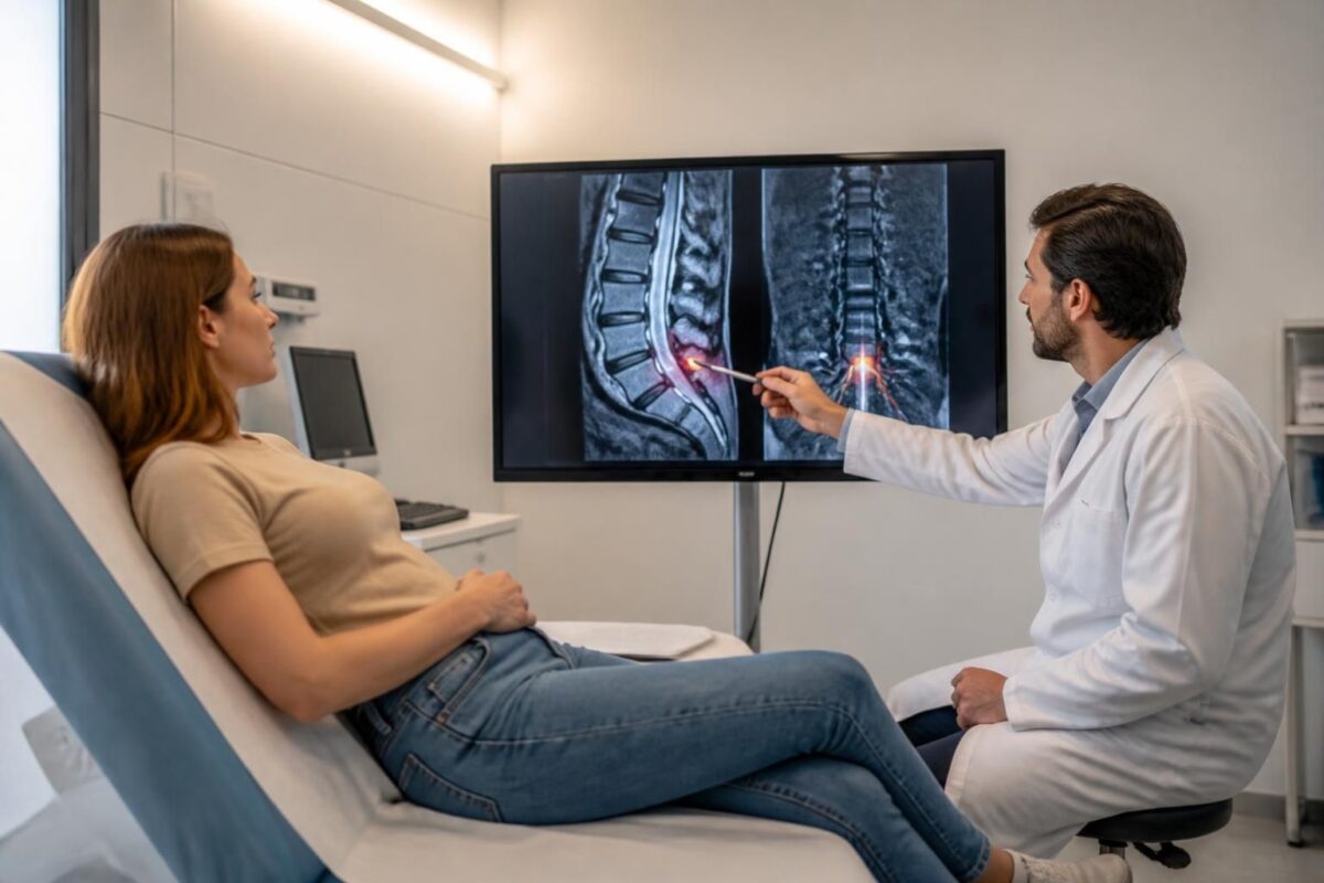
IN BRIEF
|
In brief, the MRI is the key tool for evaluating a herniated disc and sciatica. It specifies the location, type (protrusion, extrusion) and degree of compression of the nerve roots, guiding choices for non-surgical or surgical treatment. Its operation relies on multi-directional slices without radiation, allowing visualization of the disc, nerves, and signs of inflammation to facilitate diagnosis and follow-up planning. Available in specialized clinics, this examination improves the accuracy of diagnosis and management of lower back pain and sciatica. |
Summary on herniated disc and sciatica: what the MRI can show.
The MRI is the key tool for evaluating a herniated disc and sciatica. It reveals where the disc is displaced and if the nerve is compressed. It allows distinguishing three main manifestations: bulge, protrusion, and extrusion, describing the extent and severity of the hernia. The MRI specifies the affected level, the foraminal space, and can detect signs of inflammation or sequestered fragments, which helps guide non-invasive treatment options or targeted interventions.
About the author
Dr. Sylvain Desforges, B.Sc., D.O., N.D., osteopath, is the founding president of TAGMED and the ACMA association. He is devoted to managing chronic spinal pain, lower back pain, and pain associated with sciatica, especially when the pain radiates to the buttock, leg, calf, foot, or toes.
Through SOS Sciatica, he publishes educational content on the symptoms of sciatica, possible causes such as herniated disc, disc pinching, foraminal stenosis, or irritation of a lumbar nerve root, as well as non-surgical approaches that can be considered based on the evaluation. At the TAGMED Clinic, care may include, depending on the case, neurovertebral decompression, specific osteopathy, or precision impactor.
For an evaluation related to sciatica treatment in Montreal / Mont-Royal or in Terrebonne, consult the available services at the TAGMED Clinic.
This guide explains what the MRI can reveal in the case of a herniated disc associated with sciatica. It describes the typical signs observed, the different types of disc deformities, and how these images guide the choices of non-surgical management, particularly through the services of the TAGMED Clinic.
Types of deformities visible on the MRI
A disc bulge means that the disc slightly overflows beyond the vertebral border, often asymptomatic. A protrusion is a focal projection of disc material with a base wider than the dome. An extrusion occurs when disc material is projected with a narrow base and may compress the nerves. Fragments may separate (sequestration).
Signs of risk and link with sciatica
The sciatic pain follows the path of a compressed nerve root. The MRI can show nerve compression at the lumbar segments, for example, L4-L5 or L5-S1, and signs of foraminal stenosis or spinal stenosis. These indicators help the clinician evaluate non-surgical options and plan follow-up.
How the MRI helps guide treatment
The MRI does not provide a definitive diagnosis by itself. It complements the clinical evaluation and guides management decisions. Depending on the case, non-invasive approaches may be considered at the TAGMED Clinic, and certain situations may be compatible with motorized neurovertebral decompression offered by specialized care. For more information, consult the relevant resources.
To delve deeper into certain aspects, you can consult dedicated resources on muscle strength and herniated disc, or related questions on mechanical causality. Additional resources on interpreting the MRI and warning signs are available on MRI spine and on the guides provided by Sciatica and pain when straightening up.
Additional information on management and clinical scenarios is accessible through the following resources: Sos Hernie Discale and Sos Herniatedisc. For questions related to mobility and daily challenges, refer to guides on neurovertebral decompression and the options available at the TAGMED Clinic.
You can also find information on the impact of movements on sciatic pain and on difficulties in descending stairs to better understand the associated symptoms and clinical evaluation.
Medical disclaimer: The information and advice provided on this site do not replace the advice, diagnosis, or treatment of a healthcare professional. Please note that Dr. Sylvain Desforges, osteopath, is neither a doctor of medicine nor a physician, and is not a specialist in a medical specialty as defined by the College of Physicians of Quebec. Manual medicine, functional medicine, and sports medicine as described on this site exclude any treatment or medical diagnosis done by a physician or medical specialist. Always consult your doctor for any medical questions. For more details, please read our full Legal Notice.
- Precise location of the hernia: vertebral level and side.
- Type of disc projection: bulging, protrusion, or extrusion.
- Compression of a nerve root: affected root and level.
- Foraminal stenosis or spinal canal stenosis and its impact on the nerves.
- Possible sequestered disc fragment.
- Disc condition: hydration and loss of height.
- Signs of nerve irritation or compression (edema/radiculopathy).
- Adjacent structures: foramina and facet joints, possible osteoarthritis.

This sheet clearly and practically presents what an MRI can show when a disc herniation associated with sciatica is suspected. It describes the main radiological signs: location, type of disc deformation, impact on nerve roots and the spinal canal, as well as the implications for the choice of non-surgical or surgical treatments.
What is a disc herniation and how does it cause sciatica
A disc herniation occurs when the soft core of the intervertebral disc migrates through the fibrous ring and can compress a nerve root emerging at the lumbar level. This compression triggers pain that radiates to the buttock, thigh, leg, and sometimes the foot — this is referred to as sciatica. MRI is the most useful imaging tool for visualizing both the disc and nerve structures.
Herniated disc and sciatica: why an accurate diagnosis matters
IN SHORT In short, disc herniation and sciatica cause lower back pain and radicular pain. A precise diagnosis identifies the source, localizes the impairment, and guides the most appropriate treatment, avoiding unsuitable treatments. The diagnosis combines a rigorous clinical examination…
Herniated disc and sciatica: factors that influence progression
IN BRIEF In brief, disk herniation and sciatica are two distinct but often associated mechanisms that depend on a set of mechanical and inflammatory factors. The main advantage of understanding these factors is to guide an adapted and progressive rehabilitation,…
What the MRI can show
The MRI provides detailed images of soft tissues and allows for evaluation:
Location and level affected
The level of the affected disc (L4-L5, L5-S1, etc.) and the pathway of nerve involvement can be specified.
Disc condition and type of deformation
Common terms describe the displacement of disc material: protrusion (broad projection at the base), extrusion (narrower base and often pronounced nerve compression), and cases of overhanging or collapse of the disc. Fragments may be sequestered.
Compression of roots and nerve signal
The MRI can show nerve root compression and signs of nerve reaction, such as variations in T2 signal indicating inflammation or edema.
Spinal canal condition and foraminal stenosis
Narrowing of the spinal canal or foraminal escape may exist, limiting space for the roots and worsening symptoms.
Associated signals and differential diagnosis
Elements such as inflammatory anomalies, signs of targeted degeneration, or Modic changes may accompany disc involvement and guide management.
Herniated disc and sciatica: when the pain becomes persistent
IN BRIEF In short, disc herniation and sciatica refer to lower back pain radiating into the leg due to nerve compression. Follow-ups link pain pathway, neurological examination, and relevant imaging to guide management, usually without surgery. Benefits: self-management, maintenance of…

Herniated disc and sciatica: when the cause is mechanical
IN SHORT In short, Herniated disc and sciatica refer to radicular pain due to a mechanical cause when the nucleus of the disc protrudes and irritates a nerve root. The benefits of management include quick pain relief, maintenance of mobility,…
Impact on management and clinical decisions
Symptoms-imaging correspondence
The results must be interpreted alongside the clinical history and examination. Chest pain or major neurological signs require a different approach than low back pain without deficits.
When the MRI guides treatment
Depending on the location and severity of the compression, imaging may direct toward conservative care (manual therapy, exercises, pain management) or targeted options such as decompression or surgical repair when necessary.
Particular cases and follow-up
Repeated follow-ups or additional examinations may be recommended if symptoms persist or change, or if new signs appear.

Herniated disc and cauda equina syndrome: warning signs
IN BRIEF In brief, Herniated disc and cauda equina syndrome describe severe compression of the lumbar roots with warning signs requiring urgent management. The warning signs help prevent major sequelae and facilitate rapid referral to emergency services. The benefits include…

Herniated disc and muscle strength: why to test it
IN BRIEF The topic is the herniated disc and muscle strength. Testing muscle strength in this context helps assess the ability of the back and trunk muscles to support the spine and identify weaknesses that may exacerbate pain or limit…
Limits and complementarity of imaging
It is important to remember that the MRI does not explain pain on its own and that physical and psychosocial factors play a role. A comprehensive approach, combining clinical evaluation, imaging, and functional objectives, is recommended to decide on the best treatment plan.
| MRI Elements | Concise Interpretation |
|---|---|
| Disc herniation with protrusion, extrusion, or sequestration | May compress a nerve root and explain sciatica. |
| Location of the lesion and affected root (level: L4-L5, L5-S1, etc.) | Guides the painful pathway and possible deficits. |
| Foraminal stenosis (narrowing of the foramen) | May explain radicular pain and inflammation. |
| Nerve root compression | Indicates a risk of deficit and referred pain. |
| Disc sequestration | Fragment may migrate and worsen symptoms. |
| Disc degeneration with decreased T2 signal | Indicates fragility of the disc and increased risk of herniation. |
| Periradicular inflammation (edema around the root) | Contributes to pain and radicular sensitivity. |
| Absence of visible radicular anomaly | Pain may persist without identifiable compression; clinical correlation needed. |

Herniated disc and diminished reflexes: possible neurological sign
IN BRIEF In short, a herniated disc is a partial rupture of the disc ring that can compress a nerve root and cause a neurological sign such as decreased reflexes. This condition presents as pain, often in the lower back…

Herniated disc and loss of sensation: why evaluate
IN BRIEF A herniated disc is a protrusion of the gelatinous nucleus of an intervertebral disc that can compress a nerve root, causing radicular pain and a loss of sensation in an area of the body. Quickly assessing this condition…
Testimonials: what the MRI can show about disc herniation and sciatica
Marie, 42 years old, suffered from pain radiating to her buttock and leg. The MRI showed a lumbar disc herniation with a protrusion slightly compressing the nerve root. This diagnosis allowed for the planning of a non-invasive treatment focused on osteopathy and targeted exercises, and the symptoms gradually decreased over a few weeks.
Pierre, 54 years old, experienced pain with debilitating sciatica. The MRI revealed a disc herniation with extrusion, and sequestered fragments were strongly compressing the nerve. This image guided toward non-surgical alternatives and a tailored rehabilitation program that restored mobility and reduced pain.
Sophie, 29 years old, awoke with nighttime pain and tingling in her leg. The MRI highlighted a foraminal stenosis causing a narrowing of the canal and nerve pressure. The visual diagnosis was reassuring and allowed for the adaptation of treatment and muscle strengthening to improve posture and relieve symptoms.
Marc, 63 years old, has persistent low back pain radiating into his leg. The MRI showed a disc bulge without major extrusion, which was reassuring and facilitated a conservative approach: manual therapy, exercises, and work on movement habits, with progressive and lasting results.
Alice, 46 years old, has recurrent sciatica. The MRI identified an isolated disc protrusion with slight compression on the nerve root. This visual assessment guided a non-surgical management plan and sessions of osteopathy and strengthening, resulting in a notable improvement in daily life.
Disc Herniation and Sciatica: What the MRI Can Show
Dr. Sylvain Desforges, osteopath, naturopath, and manual medicine specialist, is the founding president of TAGMED clinics and the ACMA association. His career is dedicated to innovation in healthcare and the sustainable improvement of comfort and mobility for his patients. In his practice, he places a central emphasis on imaging as a tool for understanding and decision-making, particularly when it comes to disc herniation and sciatica. The goal is to establish, with the patient, an evidence-based approach to preserve or restore function, while prioritizing non-surgical options whenever possible.
When an intervertebral disc moves abnormally or deforms, magnetic resonance imaging (MRI) can reveal details that guide diagnosis and treatment planning. The MRI notably distinguishes between different types of disc pathology: bulge (or disc bulge), which extends beyond the vertebral border without definitive base loss; protrusion, focal projection of disc material with a base wider than the dome; extrusion, where disc material extends far beyond the base and can significantly compress nerve roots; and sometimes fragments that sequester, indicating instability and persistent pain. These variations have major clinical implications for sciatica and the localization of nerve compression.
Beyond disc content, the MRI allows for the evaluation of the nerve and bony environment. One can observe nerve root compression in the lumbar canal or at the foraminal level, a foraminal stenosis that can limit nerve passage and exacerbate pain radiating to the buttock, leg, calf, or foot. Signs of inflammation adjacent to the root and changes in disc signal (such as water alterations and signs of annular involvement) can guide the estimation of prognosis and the appropriateness of conservative or surgical treatment. The MRI can also highlight concomitant factors such as facet joint arthropathy or degeneration of vertebral endplates, which influence the overall approach to radicular pain.
For Dr. Desforges, interpreting the MRI is accompanied by a rigorous clinical evaluation. The association between radiological signs and the patient’s actual symptoms determines the relevance of non-invasive interventions – such as manual therapies, motorized spinal decompression, osteopathic approaches, and physically active therapies – or, when necessary, referral to more targeted options. The objective is to use the MRI as a compass that informs therapeutic choices, while remaining vigilant that radiological anomalies do not always correlate with pain or functional dissociation proportional to the imaging degree.
This framework, nourished by an evidence-based practice and technological innovation, allows for providing care tailored to each patient, prioritizing solutions that restore mobility and reduce pain related to disc herniation and sciatica. To learn more about the approach and available services, contact TAGMED Clinic.
Quick summary: this article explains what MRI can reveal in cases of disc herniation and sciatica, describing the various patterns of disc deformity and their significance for pain and treatment, while indicating how to interpret the results in relation to clinical evaluation.
On lumbar MRI, several patterns of intervertebral disc deformation and their impact on nerve roots can be distinguished. The bulge corresponds to a general protrusion beyond the vertebral border, the protrusion is a focal projection with a base wider than the dome, and the extrusion involves a narrow material projection at the base with an increased risk of nerve compression, sometimes with sequestered fragments. These configurations influence the degree of nerve compression and guide therapeutic decisions based on clinical assessment.
The precise location of the affected disc and its effect on nerve roots allow for anticipating clinical manifestations such as sciatica, thigh pain, or sensory symptoms in the leg. Foraminal involvement may lead to compression of foraminal roots, while foraminal or central stenosis may limit available space for the nerves and influence the treatment plan.
In addition to disc patterns, the MRI can reveal other important elements such as foraminal or central stenosis, signs of inflammation around the nerve, and Modic changes of the vertebral endplates, which may accompany chronic pain. One must also be vigilant regarding the potential diagnosis of cauda equina syndrome, which constitutes a medical emergency.
However, the MRI is not synonymous with pain: certain abnormalities may be present in asymptomatic patients and vice versa. Interpretation should be performed in correlation with the clinical examination and the patient’s history. Radiological images may guide towards non-surgical approaches or, conversely, towards a surgical evaluation when neurovascular signs progress or when a persistent deficit remains despite treatment.
For further insights into the practical aspects and clinical implications, refer to specialized resources such as diagnosis of disc hernias and MRI alert and Disc hernia, sciatica and cruralgia.
For additional information related to symptoms and their effects on walking or limb sensations, you may refer to sciatica and heavy legs while walking, MRI and disc herniation or persistent pain in the heel.

Dr Sylvain Desforges, B.Sc., D.O., N.D., osteopath, is the founding president of TAGMED and of the ACMA association. He dedicates his practice to chronic spinal pain, low back pain and symptoms compatible with sciatica, particularly when pain radiates toward the buttock, leg, calf, foot or toes.
Through SOS Sciatique, he publishes educational content on sciatica symptoms, possible causes such as disc herniation, disc narrowing, foraminal stenosis or irritation of a lumbar nerve root, as well as non-surgical approaches that may be considered according to the evaluation. At Clinique TAGMED, care may include, depending on the case, motorized neurovertebral decompression, specific osteopathic care or precision percussion.
For an evaluation related to sciatica treatment in Montreal / Mount Royal or sciatica treatment in Terrebonne, please consult the services available at Clinique TAGMED.
Medical disclaimer
The information published on SOS Sciatique is provided for educational purposes only and does not replace a consultation, diagnosis, opinion or treatment provided by a physician or another authorized healthcare professional. The author of this article is neither a medical doctor nor a specialist in a medical specialty recognized by the Collège des médecins du Québec. The osteopathic, manual medicine or functional support approaches described on this site do not constitute medical acts and are not intended to replace medical follow-up.
Consult a physician or emergency service in cases of loss of bladder or bowel control, anesthesia in the saddle area, significant or progressive weakness in the leg or foot, fever, recent trauma, severe pain or rapid worsening of symptoms. For more details, please read our Legal Notice.