

Sciatica and pain when straightening up
22/09/2026
Sciatica and stairs: when movement worsens pain
22/09/2026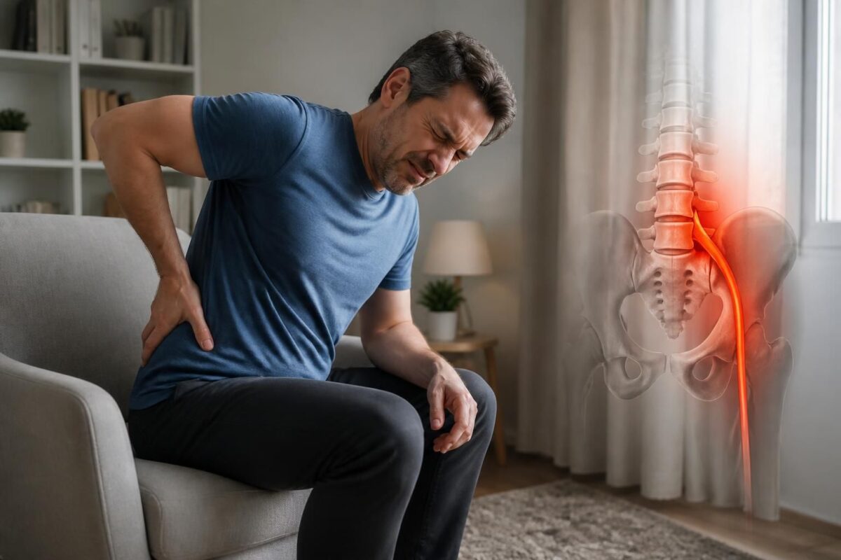
IN BRIEF
| In short, disc herniation and sciatica refer to lower back pain radiating into the leg due to nerve compression. Follow-ups link pain pathway, neurological examination, and relevant imaging to guide management, usually without surgery. Benefits: self-management, maintenance of daily activities, durable pain reduction, and functional improvement through conservative treatments and physiotherapy; injections or surgery remain reserved based on progression and neurological signs. In Montreal, this service offers quick access and sustainable improvement while avoiding inactivity. |
Disc Herniation and Sciatica: When Pain Becomes Persistent
The disc herniation can compress a nerve root and cause pain radiating into the leg, often referred to as sciatica.
A pain that persists does not necessarily mean the herniation is worsening: factors such as nerve sensitivity, deconditioning, sleep, and fear of movement can sustain symptoms even if anatomy changes.
Imaging can locate the injury, but the presence of a herniation alone does not explain the symptoms: it is important to connect the level, side, and clinical picture.
- Indicator 1: some herniations decrease over time, but recovery can be slow.
- Indicator 2: persistent pain warrants a reevaluation of the diagnosis and functional objectives.
- Indicator 3: chronicity does not mean the pain is imaginary; it results from biological, mechanical, and contextual factors.
For management, it is necessary to stay active and maintain normal activities as much as possible, with adaptations as needed. Treatment is usually conservative (pain relief, physiotherapy, exercises), and injections may be considered if necessary. Surgery is only considered in the case of a neurological emergency or treatment failure after several weeks.
Warning signs requiring prompt consultation: new or progressive weakness, foot drop, urinary/fecal issues, saddle numbness, fever, or major trauma.
Quick summary: sciatica pain can become persistent without the disc herniation necessarily continuing to grow. The crux is that nerve sensitivity, deconditioning, sleep, fear of movement, and other factors can maintain symptoms. Effective evaluation combines symptoms, function, neurological examination, and imaging when relevant, aiming to guide appropriate management without relying solely on imaging.
For more information, you can consult additional resources on the following links: Disc herniation and pain without back pain: is it possible, or Disc herniation and driving.
Understanding the Link Between Disc Herniation and Sciatica
A disc herniation can compress a nerve root and cause pain that radiates into the buttock, leg, or foot. However, the presence of a herniation visible on imaging does not solely explain the symptoms. Factors such as increased nerve sensitivity, lack of muscular conditioning, sleep disturbances, and fear of movement can sustain pain even when anatomy changes.
The pain pathway, motor and sensory signs, and functional adaptation must be consistent with the location and level of the herniation observed in imaging when performed.
Essential Clinical Indicators
Indicator 1: some herniations diminish over time, but recovery can be slow and incomplete.
Indicator 2: persistent pain prompts reevaluation of the diagnosis and functional goals to redirect the treatment plan.
Indicator 3: chronicity does not mean the pain is imagined; it often reflects multiple biological, mechanical, and contextual factors.
Assessment Elements and Clinical Concordance
The diagnosis relies on the concordance between pain pathway, muscle strength, sensitivity, reflexes, and function. Imaging should be interpreted in relation to the clinical picture and not as isolated proof of a cause.
1 Pathway: precisely locate pain, paresthesias, and areas of numbness.
2 Strength and function: assess key movements, gait, and tasks revealing potential weakness.
3 Imaging: connect the level and side of disc morphology to the clinical picture rather than concluding a single cause.
4 Progression: monitor the trend of symptoms and define criteria that may alter the therapeutic direction.
Stay Active and Targeted Imaging
Clinical recommendations encourage self-management and maintaining normal activities when possible, with adaptations according to abilities. Imaging should only be considered if it can modify management. Avoiding inappropriate movements or exercises during an acute episode is a cautious approach when symptoms are complex.
For further exploration, additional resources on nerve pathways and diagnosis can be found at Disc herniation and pain without back pain: is it possible.
Management and Treatment Options
In Montreal and Mont-Royal, the TAGMED Clinic offers tailored solutions case by case, with non-surgical options preferred when possible. For more information on available options, visit TAGMED Clinic.
Additional information on neurovertebral decompression approaches and other related options can be viewed at neurovertebral decompression.
When to consider a specialized evaluation
If there is a progressive motor deficit, loss of perineal sensation, marked bilateral pain, or urinary or fecal disturbances, urgent care or a specialized consultation is necessary. Persistent pain that does not improve functionally may warrant reevaluation and discussion of the following options, while avoiding inappropriate treatments if they do not demonstrate effectiveness.
For additional resources, you can consult Inflammation of the nerve root and Paramedial disc herniation and sciatica.
When to seek prompt or emergency care
Signs requiring quick assessment: new, significant, or rapidly progressive weakness; recent foot drop; deteriorating gait; urinary or intestinal disturbances; numbness of the saddle or perineum; significant bilateral symptoms or fever; major trauma or general deterioration.
In these cases, do not wait for a routine appointment. Prompt care can influence the prognosis.
References and additional resources
For detailed information on trajectories and mechanisms, see the following specialized resources: Disc herniation and pain without back pain, is it possible, Driving and symptom worsening, Inflammation of the nerve root, Possible painful path.
For general information on treatments and outcomes, refer to Disc herniation and pain without back pain, is it possible.
Additional information on approaches and management options can be found at Herniated disc and Herniated disc and sciatica.
For general resources on the nerve path and healing, see Path of the nerve.
Further information can be found on Disc herniation and pain without back pain and Lumbar osteoarthritis and sciatica.
These references are provided for informational purposes and do not replace a medical consultation.
For any additional information on diagnostics and provided treatments, visit TAGMED Clinic and neurovertebral decompression.
Medical disclaimer: The information and advice provided on this site do not replace the opinion, diagnosis, or treatment by a healthcare professional. Please note that Dr. Sylvain Desforges, osteopath, is neither a physician nor a medical doctor, and is not a specialist in any medical specialty as defined by the Collège des médecins du Québec. Manual therapy, functional medicine, and sports medicine as described on this site exclude any treatment or diagnosis made by a physician or medical specialist. Always consult your doctor for any medical questions. For more details, please read our complete Legal Notice.
- Analysis axis: concordance between symptoms, examination, and imaging to guide management.
- Persistence ≥6 weeks without clear improvement may reflect biological, mechanical, and contextual factors.
- Chronicity: does not necessarily mean that pain is imaginary; it often results from multiple interactions.
- Urgency: the onset of progressive weakness, foot drop, or cauda equina signs requires rapid assessment.
- Pain pathway and motor or sensory signs help refine diagnosis and priorities.
- Path and function: accurately locate pain, paresthesias, and numbness; assess strength and gait.
- Targeted imaging: useful when the result can change management; not systematic.
- Non-surgical management: maintain appropriate activity, physiotherapy, and strengthening; avoid prolonged inactivity.
- Traction and decompression: traction is not recommended as a standard treatment; motorized decompression requires information and caution.
- Monitoring: track evolution and establish redirection criteria if symptoms persist or worsen.
- Quick consultation: alerts to recognize: urinary or fecal disturbances, fever, major trauma, significant bilateral pain.

This practical summary explains when a herniated disc and sciatica become persistent, and which steps to prioritize to guide management without delaying the return to activity. The goal is to help discern what relates to natural evolution, what requires reevaluation, and which options to discuss to limit functional and painful impact.

Herniated disc and sciatica: when the cause is mechanical
IN SHORT In short, Herniated disc and sciatica refer to radicular pain due to a mechanical cause when the nucleus of the disc protrudes and irritates a nerve root. The benefits of management include quick pain relief, maintenance of mobility,…
When pain becomes persistent
A persistent sciatic pain does not necessarily indicate that the disc herniation has worsened. Factors such as nerve sensitization, deconditioning, sleep, and fear of movement can maintain symptoms even if the anatomy does not evolve favorably. It is crucial to regularly assess functional impairment and response to treatment, rather than relying solely on imaging.

Herniated disc and cauda equina syndrome: warning signs
IN BRIEF In brief, Herniated disc and cauda equina syndrome describe severe compression of the lumbar roots with warning signs requiring urgent management. The warning signs help prevent major sequelae and facilitate rapid referral to emergency services. The benefits include…
Assessment and clinical concordance
The diagnosis relies on the agreement between symptoms, function, neurological examination, and imaging when relevant. A visible herniation on an MRI does not alone mean it is the cause of pain: the concordance between the level, side, and clinical picture is decisive. The evolution of motor and sensory signs guides evaluation priorities and therapeutic decisions.

Herniated disc and muscle strength: why to test it
IN BRIEF The topic is the herniated disc and muscle strength. Testing muscle strength in this context helps assess the ability of the back and trunk muscles to support the spine and identify weaknesses that may exacerbate pain or limit…
Key points to guide management
Point 1
Some hernias decrease over time, but symptom recovery can be slow. Patience and the maintenance of a suitable active program often remain effective during the natural resolution phase.
Point 2
A pain that persists beyond the initial period deserves a reevaluation of the working diagnosis and functional goals to adjust treatments and activities.
Point 3
Chronicity does not mean the pain is imaginary; it often reflects an interaction of biological, mechanical, and contextual factors. A multimodal and progressive approach is generally preferred.

Herniated disc and diminished reflexes: possible neurological sign
IN BRIEF In short, a herniated disc is a partial rupture of the disc ring that can compress a nerve root and cause a neurological sign such as decreased reflexes. This condition presents as pain, often in the lower back…
Elements of clinical evaluation
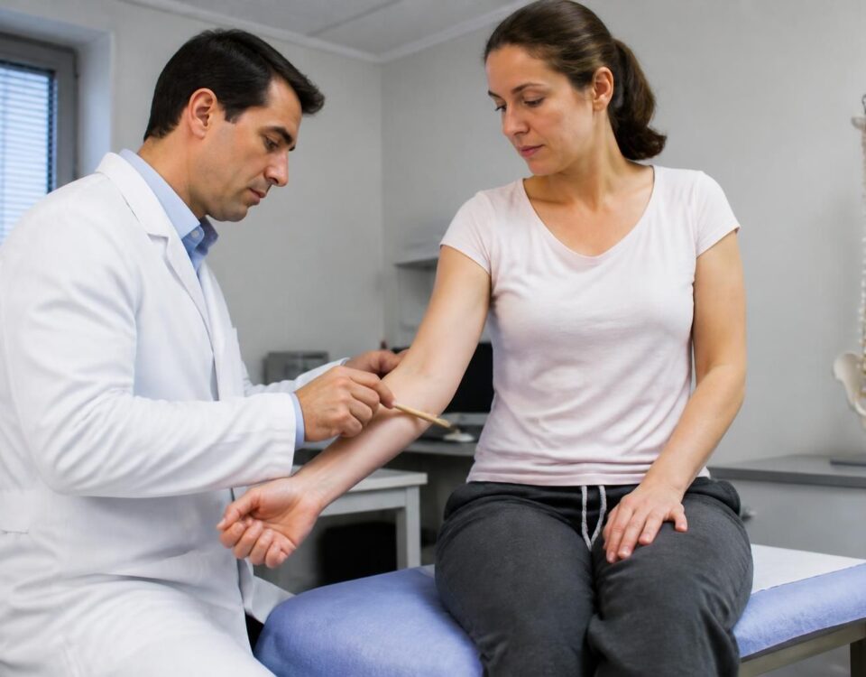
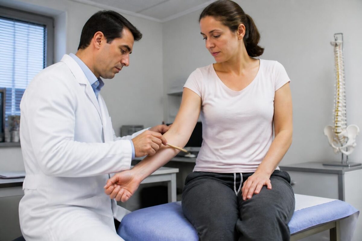
The painful path is compared to motor, sensory, and functional signs. The examination aims for concordance between what sources the pain and what tone, strength, sensitivity, reflexes, and functional autonomy show.
1 Path
Precisely localize the pain, paresthesias, and numbness areas to relate the painful territory to a possible nerve root.
2 Strength and function
Compare key movements, evaluate walking and tasks that reveal potential weakness to guide rehabilitation priorities.
3 Imaging
Relate the level and side of the lesion to the clinical examination rather than stopping at an isolated report. Imaging can complement evaluation but should not be interpreted in isolation.
4 Evolution
Measure the trend of symptoms and define criteria for redirecting treatment if necessary.
Herniated disc and loss of sensation: why evaluate
IN BRIEF A herniated disc is a protrusion of the gelatinous nucleus of an intervertebral disc that can compress a nerve root, causing radicular pain and a loss of sensation in an area of the body. Quickly assessing this condition…
Practical management and recommendations
Staying active is preferable when possible. The goal is self-management and maintaining normal activities, adjusting efforts according to capabilities and body signals. Targeted imaging is useful only if it can change management. Lumbar traction is not recommended as standard treatment; any motorized decompression should be discussed transparently regarding the uncertainties of the data.

Herniated disc and foot pain: L5 or S1?
IN BRIEF In brief, Disc herniation and foot pain related to the L5-S1 level explain how a hernia can irradiate towards the foot and how to determine if the pain originates from L5 or S1. This approach aims for a…
When to seek prompt consultation
Consult urgently or quickly in the presence of new and significant weakness, foot drop, worsening walking, urinary or bowel problems, numbness of the saddle or perineum, or significant bilateral symptoms accompanied by fever, major trauma, or general deterioration.
Herniated disc and spondylolisthesis: possible nerve irritation
IN BRIEF The lumbar disc herniation and spondylolisthesis refer to a possible compression of the nerve roots leading to sciatic pain and sometimes paresthesia or muscle weakness. This nerve irritation can mostly be managed through conservative measures and medication, providing…
Key points to remember
A lasting improvement in function is more useful than mere reduction in pain. A sciatic pain possibly related to a disc herniation requires a consultation to verify concordance between symptoms, examination, and imaging and to discuss management options. Persistence after several weeks does not exclude future improvement and does not necessarily require repeated MRI, unless it could influence the clinical decision.
| Aspect | Impact / Concise description |
|---|---|
| Definition and link | The disc herniation does not explain everything: pain can persist even when anatomy is stable and the clinical picture is coherent. |
| Persistence | Persistence > 6 weeks or months requires reevaluation of the diagnosis and functional goals. |
| Role of imaging | Imaging must be interpreted in relation to clinical signs; a visible herniation does not necessarily imply symptoms. |
| Clinical evaluation | The examination looks for concordance between the pain path, strength, sensation, and function. |
| Maintenance factors | Biological and mechanical factors like nerve sensitivity, deconditioning, sleep, and fear of movement can maintain pain. |
| Self-management and activity | Staying active and adapting activity is recommended to limit inactivity and promote recovery. |
| Targeted imaging | Consider targeted imaging only if the result would change management. |
| Urgency and signs to monitor | Quick consultation in the presence of new weakness, urinary issues, significant bilateral pain, fever, or trauma. |
| Prognosis and goals | Chronicity does not prevent improvement; the goal is functional progress through rehabilitation and tailored therapies. |

Herniated disc and lumbar osteoarthritis: combined causes of sciatica
IN BRIEF The subject is the association between herniated disc and lumbar osteoarthritis as combined causes of sciatica and femoral neuralgia. This interaction explains the low back pain and nerve irradiations. Benefits and applications: allows differentiation of pain related to…
Testimonies on disc herniation and sciatica: when pain becomes persistent
Marie, 42 years old — “My lower back pain became persistent after months of diffuse pain. The feeling of radiating into my buttock and leg was constant, and my sleep suffered. What I learned is that the disc herniation does not explain everything: you need to look at the pain path, neuro-muscular state, and lifestyle habits. Thanks to a comprehensive assessment and a tailored program, the pain gradually decreased, and I was able to return to my daily activities without fearing every movement.”
Jean, 55 years old — “After several months without improvement, I realized that seeing an image on the MRI was not enough to explain everything. My doctor evaluated the pain pathway, strength, and function, then adjusted the treatment accordingly. The plan focused on physical activity and targeted exercises, rather than corticosteroids or medications alone. Today, the sciatica is much less present and I have returned to my daily tasks with confidence.”
Sophie, 33 years old — “The night pain was unbearable and my sleep had been disrupted for too long. I received a reevaluation that placed clinical assessment at the heart of the diagnosis and highlighted that persistent pain could be fueled by fear of movement and deconditioning. By working on simple exercises, mobility and motor control improved, and sleep became restorative again.”
Marc, 50 years old — “I started to notice weakness in my foot and a gradually deteriorating walk. The path was clear: we had to quickly redirect the assessment towards appropriate management, prioritizing the maintenance of suitable activity and stability exercises. The process allowed me to keep hope and limit periods of inactivity.”
Claire, 46 years old — “After months of pain, I understood that persistence did not necessarily imply a herniated disc progression, but often involved several biological and mechanical factors. The support emphasized education, maintaining activity, and functional adjustments. My daily life has approached that of a normal life, with less pain and more mobility.”
Luc, 39 years old — “I discovered that imaging could show a disc herniation in people without pain, which reassured me about the importance of a solid clinical link. The team took the time to examine the pain pathway, sensitivity, and strength, rather than treating an isolated aspect of the problem. In short, we worked on balancing activity, pain, and function, and I regained the ability to do what I love without fear.”
Dr. Sylvain Desforges is an osteopath specializing in the management of chronic pain and a pioneer in integrating manual techniques and complementary medicine. As the founding president of TAGMED clinics and the ACMA association, he combines scientific rigor with a human approach to guide his patients towards long-term improvement in their quality of life. His area of expertise particularly covers spinal pain and radicular manifestations related to a disc herniation and sciatica, prioritizing evidence-based interventions and transparent communication about available options.
In this context, he emphasizes that the presence of a disc herniation does not necessarily mean that the pain will continue to progress or that the current anatomy is the sole cause of the symptoms. Several factors can maintain persistent pain, such as increased nerve sensitivity, muscle deconditioning, sleep disturbances, and fear of movement. The goal is to assess not only the level of impairment but also the functional impact and the patient’s ability to maintain or regain their daily activities.
The diagnostic approach proposed by Dr. Desforges closely links symptoms to function, combining neurological examination and, when relevant, imaging. The objective is to construct a clear synthesis between the pain pathway, motor and sensory signs, and radiological elements. This approach avoids hasty interpretations and helps prevent unnecessary treatments based solely on an image.
Marker 1, Marker 2, and Marker 3 outline a useful progression to understand the dynamics of pain. Marker 1 indicates that some hernias may decrease over time, but recovery can be slow. Marker 2 emphasizes that persistent pain warrants a reevaluation of the initial diagnosis and functional objectives. Marker 3 reminds that chronicity is not synonymous with imagined pain: it often reflects the interaction of biological, mechanical, and contextual factors that require a reassessed and personalized approach.
The pain pathway is then matched with the patient’s motor signs, sensory signs, and functional abilities. The clinician precisely localizes the pain and paresthesias, assesses the strength and function of key movements, and examines walking and tasks that reveal potential weaknesses. Imaging is useful as a complement when the level and side of the impairment correspond to the clinical picture, but should not be interpreted in isolation. Finally, the evolution of symptoms is measured to define the criteria that may guide towards another management approach.
Staying active remains a central recommendation. Contemporary guidelines prioritize self-management and maintaining normal activities as much as possible, with adaptations suited to the patient’s capacity. Targeted imaging is considered when the results can genuinely modify the treatment plan. Conversely, approaches such as traction are not recommended as standard treatment for low back pain with or without sciatica, and any motorized decompression must be discussed transparently regarding the uncertainty of the data.
There are signs that necessitate prompt consultation or emergency care. New and progressive weakness, a foot drop, new urinary or bowel disturbances, numbness of the saddle or perineum, significant bilateral symptoms, fever, major trauma, or general deterioration require quick evaluation and should not wait for a routine appointment.
In practice, a lasting improvement in function is often more significant than a simple one-off reduction in pain. If you are suffering from low back pain associated with sciatica, a specialized consultation can help clarify the alignment between symptoms, examination, and imaging, and consider management options tailored to your situation.
In short, a disc herniation associated with sciatica can become persistent without the “anatomy” necessarily evolving in an alarming manner. The key message is that persistent pain often results from a combination of nerve sensitivity, deconditioning, sleep, and psychological or contextual factors, rather than a purely structural mechanism. A function-based, activity-oriented, and rigorously clinical evaluation approach allows for effective and personalized treatment guidance.
The diagnosis of low back pain with radiculopathy relies on a concordance between pain pathway, motor function, and examinations, rather than solely on imaging. A disc herniation visible on MRI is only relevant if the level, side, and clinical picture align. This vigilance prevents hasty conclusions and guides towards care that accurately reflects the patient’s reality.
Chronicity does not mean that pain is undeserved or inevitably linked to anatomical progression. It often reflects an interaction between biological, mechanical, and contextual factors, including nerve sensitivity and overall functional performance. Understanding this landscape allows for targeting functional goals and readjusting daily activities without guilt or prolonged immobilization.
Clear guidelines guide management: some hernias decrease over time, but symptom recovery may be slower, and persistent pain justifies a reevaluation of the working diagnosis and functional goals. A key concern remains: chronic pain does not stop at a simple pain/imaging relationship; it requires a multidisciplinary approach tailored to each patient’s needs.
The pain trajectory must be interpreted with motor, sensory, and functional signs. The clinical examination seeks to establish a concordance between pain location, muscle strength, reflexes, and functional capabilities, in order to determine whether the symptoms stem from a specific disc level or other musculoskeletal mechanisms.
In practice, current recommendations encourage maintaining normal activity as much as possible and appropriate self-management. Imaging is useful when results could modify a therapeutic decision, but it should not be used as the sole diagnostic tool. Traction or repeated approaches without solid indication do not replace a plan centered on appropriate decompression and functional rehabilitation. For resistant cases, options such as epidural injection or other targeted interventions may be considered after a thorough discussion of the benefits and uncertainties.
When to quickly seek specialized evaluation? In the presence of new or progressive weakness, a foot drop or deterioration in walking, urinary or fecal signs, saddle anesthesia, or any febrile, traumatic, or generalized signs; these elements require urgent management and do not substitute for a planned consultation.
For patients concerned with sciatica associated with a disc herniation, a specialized consultation allows for checking the concordance between symptoms, examination, and imaging, and clarifying the signs to watch for as well as appropriate management options. The outlook is optimistic: even after several months, improvement is possible through targeted rehabilitation and a gradual resumption of adapted activities.
To delve deeper, you can consult additional resources that address symptoms, the role of imaging, and management options: clinical significance of extruded disc herniation, disc herniation and chronic sciatica, neurovertebral decompression and recovery after recurrent disc herniation. For complementary perspectives, also see general sources on sciatic pain and associated MRI.

Dr Sylvain Desforges, B.Sc., D.O., N.D., osteopath, is the founding president of TAGMED and of the ACMA association. He dedicates his practice to chronic spinal pain, low back pain and symptoms compatible with sciatica, particularly when pain radiates toward the buttock, leg, calf, foot or toes.
Through SOS Sciatique, he publishes educational content on sciatica symptoms, possible causes such as disc herniation, disc narrowing, foraminal stenosis or irritation of a lumbar nerve root, as well as non-surgical approaches that may be considered according to the evaluation. At Clinique TAGMED, care may include, depending on the case, motorized neurovertebral decompression, specific osteopathic care or precision percussion.
For an evaluation related to sciatica treatment in Montreal / Mount Royal or sciatica treatment in Terrebonne, please consult the services available at Clinique TAGMED.
Medical disclaimer
The information published on SOS Sciatique is provided for educational purposes only and does not replace a consultation, diagnosis, opinion or treatment provided by a physician or another authorized healthcare professional. The author of this article is neither a medical doctor nor a specialist in a medical specialty recognized by the Collège des médecins du Québec. The osteopathic, manual medicine or functional support approaches described on this site do not constitute medical acts and are not intended to replace medical follow-up.
Consult a physician or emergency service in cases of loss of bladder or bowel control, anesthesia in the saddle area, significant or progressive weakness in the leg or foot, fever, recent trauma, severe pain or rapid worsening of symptoms. For more details, please read our Legal Notice.