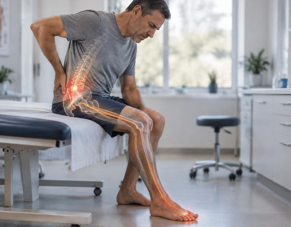



Sciatica and difficulty standing upright
17/09/2026
Sciatica and slow walking: better tolerated or not?
17/09/2026
IN BRIEF
| A herniated disc is a protrusion of the gelatinous nucleus of an intervertebral disc that can compress a nerve root, causing radicular pain and a loss of sensation in an area of the body. Quickly assessing this condition allows for the identification of emergency signs (progressive motor deficit or cauda equina syndrome), guiding an appropriate treatment and improving recovery chances by avoiding delays. The process generally involves a clinical examination and, if necessary, appropriate imaging (lumbar MRI) to locate the hernia and direct management, with concrete recommendations for prevention and recovery. In Montreal, this service is particularly recommended for rapid assessment and referral to spine specialists to ensure effective and personalized management. |
The loss of sensation related to a herniated disc indicates damage to a nerve root and deserves a quick evaluation. This assessment allows for understanding the affected level, the extent of the deficit, and the risks of neurological complications.
- Precisely locate the affected nerve and correlate sensory symptoms to the corresponding territory.
- Detect signs of neurological emergency (e.g., cauda equina syndrome, progressive motor deficit, urinary disorders).
- Determine the appropriate treatment between conservative management and surgery.
- Order appropriate examinations (notably the MRI) and, if necessary, additional tests such as EMG.
- Plan for recovery and prevent recurrences through rehabilitation and muscle strengthening.
This text explains why it is crucial to evaluate a loss of sensation associated with a herniated disc. It describes possible mechanisms, warning signs, assessment tools, and management options, referring to specialized structures like the TAGMED Clinic for appropriate evaluation and treatment.
Understanding the Link Between Herniated Disc and Loss of Sensation
A herniated disc occurs when the gelatinous nucleus of a disc protrudes from its outer layer and can compress a nerve root. This compression can cause loss of sensation or abnormal sensations in the territory supplied by the injured nerve. The typical deployment of sensory disorders depends on the affected level:
L3-L4 can cause crural involvement with decreased sensation on the anterior thigh and associated knee weakness; L4-L5 and L5-S1 can lead to sciatic radiculopathy with numbness or paresthesia along the buttock, leg, and foot. To better understand the nerve mechanisms involved, you can consult specialized resources on nerve root inflammation and disc interactions on educational websites of the network. For example, nerve root inflammation can aggravate sensory symptoms when the root is irritated by the disc protrusion.
Warning Signs and Why Evaluation is Essential
Signs Requiring Urgent Management
The cauda equina syndrome is a neurological emergency. It manifests as a loss of perineal sensation, urinary or fecal incontinence, and urinary retention. If these signs appear, emergency services must be contacted immediately, as every hour counts to preserve continence and nerve function.
Signs of Progression or Motor Deficit
A progressive motor deficit, such as the inability to lift the foot (foot drop) or worsening weakness, requires rapid neurosurgical evaluation, usually within 24 to 48 hours. A persistent motor deficit can impact recovery prognosis.
Evaluation Process
The evaluation is based on a thorough clinical examination and imaging studies. Lumbar MRI is the reference examination for locating the hernia, estimating the fragment’s volume, and checking for radicular compression. A CT scan may be used when MRI is contraindicated. Electrodiagnostic tests can be useful in some cases to assess nerve root suffering and guide prognosis.
It should be kept in mind that visible disc hernias on imaging are not always the source of symptoms. About 30% of asymptomatic individuals show a herniated disc on imaging. It is the association between the clinical picture and imaging results that guides the therapeutic decision. For more technical aspects, see the resource dedicated to MRI and diagnosis of herniated discs and radiological assessment.
Treatment Options and Follow-Up
In most cases, the herniated disc regresses spontaneously, and sensory symptoms improve over time. Initial treatment aims to relieve pain and support nerve recovery while avoiding actions that worsen pain. It may include analgesic and anti-inflammatory measures, short-term corticosteroids for intense radicular pain, and a gradual return to suitable activities. Evaluation determines if more targeted intervention is necessary based on progress.
For resistant cases or when the pain is disabling, complementary options exist. Epidural injections may be considered when medical treatment is insufficient to relieve radicular pain and help delay surgical intervention.
Surgery is considered in well-defined indications, particularly in cases of neurological emergency (cauda equina) or with a progressive motor deficit despite optimized treatment over several weeks. Microdiscectomy is the reference procedure when surgery is indicated, and it can provide rapid relief of radicular pain in the majority of cases. The outcome depends on whether the situation is urgent or not, and the overall clinical context.
For patients referred to specialized care in Montreal or Terrebonne, the TAGMED Clinic offers tailored evaluations and pathways. Consult TAGMED to make an appointment and discuss suitable management options for your situation.
Comparative table of therapeutic options
| Therapy | Common indications | Elements of typical outcomes |
|---|---|---|
| Conservative medical treatment | Radicular pain, without neurological urgency | 80% of cases progress to spontaneous improvement within 6-12 weeks |
| Epidural injections | Pain resistant to oral treatment | Significant reduction in pain; may avoid surgery in 60-70% of cases |
| Microdiscectomy | Hyperalgesic sciatica resistant after adequate treatment | Immediate relief of radicular pain in 90-95% of cases |
Key elements and practical guidance
The loss of sensation associated with a herniated disc requires a rapid and coordinated assessment. The evaluation combines a clinical examination and imaging tests to target the affected nerve root and determine the best follow-up plan. In cases where intervention is necessary, surgery is reserved for specific situations and patients who respond poorly to conservative treatment.
For consultations and management options, you can refer to the following resources, which cover all aspects of diagnosis and management, and remain within the recommended fields:
- Nerve root inflammation
- Recurrence and possible factors
- Foot pain and L5/S1 root
- Toe pain and L5 root
- Symptoms to recognize
- Herniated disc – MSD Manuals
- Herniated disc – patient education
- Spondylolisthesis and nerve irritation
For care at the TAGMED Clinic, prioritize an initial local assessment coordinated with credible specialists. TAGMED Clinic offers services tailored in Montreal and Terrebonne.
“Medical disclaimer: The information and advice provided on this site do not replace the opinion, diagnosis, or treatment of a healthcare professional. Please note that Dr. Sylvain Desforges, osteopath, is neither a medical doctor nor a physician, and is not a specialist in a medical specialty as defined by the Collège des médecins du Québec. Manual medicine, functional medicine, and sports medicine as described on this site exclude any medical treatment or diagnosis made by a doctor or medical specialist. Always consult your doctor for any medical questions. For more details, please read our full Legal Notice.”
- Sensitivity and nerve territory: loss of sensation and paresthesia in the pathway of the affected root.
- Level and location: determine the affected disc (L3-L4, L4-L5, L5-S1) to target the assessment.
- Motor deficit: weakness of muscles innervated by the affected root (e.g., tibialis anterior).
- Signs of emergency: loss of bladder/bowel control or cauda equina syndrome — act urgently.
- Evolution: persistence or worsening beyond 6 weeks justifies reassessment and imaging.
- Diagnostic orientation: correlation between symptoms and imaging to confirm a herniated disc.
- Therapeutic decision: direct towards physiotherapy, injections, or surgery based on the assessment.
- Prevention of complications: manage pain and prevent atrophy and motor deficits.
- Follow-up plan: rehabilitation and functional follow-up to optimize recovery.
- Emergencies and quick action: know how to recognize alerts and direct towards emergencies when necessary.

Quick summary
This article explains why to evaluate a herniated disc in the presence of a loss of sensation, what this loss may reveal, useful examinations, and management options. The goal is to quickly identify signs that necessitate appropriate action to limit complications and optimize recovery.
Understanding the link between herniated disc and loss of sensation
A herniated disc occurs when the gelatinous nucleus of an intervertebral disc pushes through a tear in the outer ring and can compress a nerve root. This compression often manifests as radiating pain and may be accompanied by a loss of sensation in the area served by the affected root. Depending on the level, the affected areas differ: L3-L4 may cause a loss of sensation on the anterior thigh, L4-L5 and L5-S1 lead to radicular pain and sensory disturbances in the leg and foot, and L5-S1 may affect the heel and outer edge of the foot. The loss of sensation may manifest as numbness, tingling, decreased sensitivity to touch, or altered thermal sensation.
Why evaluate quickly
Quick assessment allows for distinguishing a sensory loss related to a disk hernia from other causes and identifying signs that may require urgent management. Early evaluation also helps differentiate non-surgical treatment options from situations that may progress to intervention. In practice, a good blood, neurological, and radiological assessment guides the decision between conservative treatment, injections, or surgery when necessary, and helps prevent irreversible nerve damage.
Warning signs that require urgent consultation
Certain situations require immediate medical attention. In the presence of a sensitivity loss associated with severe pain, one should consider urgent consultation if signs such as perineal anesthesia (loss of sensitivity around the saddle area), urinary or fecal incontinence or any onset of progressive motor deficit occur. The clinical picture may suggest a cauda equina syndrome, a critical medical condition where every hour counts and surgery may be necessary to preserve continence and motor function.
How to assess: steps and examinations
Clinical interview and neurological examination
The assessment begins with the history of symptoms and a thorough neurological examination. Sensitivity loss is located, muscle strength, reflexes, and sensation to touch are assessed to determine the affected disk and root and evaluate the degree of nerve compromise.
Imaging and additional tests
The lumbar MRI is the reference examination to visualize the affected disk, the exact level, and root compression. A CT scan may be considered if an MRI is contraindicated. X-rays are of limited use in diagnosing a disk hernia but may inform the condition of the vertebrae. Electrodiagnostic tests (EMG and nerve conduction study) complete the evaluation when the diagnosis is uncertain or to estimate the prognosis for recovery.
Role of EMG and nerve conduction tests
EMG and nerve conduction tests can help confirm root involvement and guide therapeutic decision-making, especially when sensory symptoms remain more pronounced compared to imaging or when they influence postoperative prognosis.
What to do in the meantime and management options
Initially, conservative treatment aims to control pain and promote the resorption of herniated material while maintaining appropriate physical activity. Measures may include pain relievers and anti-inflammatories, gentle exercises, and physical therapy focused on strengthening stability and posture rehabilitation. When pain is resistant, alternatives such as epidural injections may be considered under radiological control to reduce inflammation and radicular pain. Persistent sensitivity loss despite conservative treatment justifies a specialized reevaluation to clarify possible indications for surgical intervention, especially when nerve compression affects function or when neurological signs progress.
| Element to evaluate | Concise justification |
|---|---|
| Location of loss | Identify the level and affected root, to guide diagnosis and therapeutic choices. |
| Associated symptoms and irradiation | Link pain, paresthesia, and nerve pathways to estimate extent and differentiate between sciatica and femoral pain. |
| Potential motor deficit | Indicates a neuronal risk and may require specialized evaluation if progression occurs. |
| Urgent cauda equina | Loss of bowel/bladder control and perineal anesthesia require rapid management (emergency). |
| Neurological risk | Progressive motor deficit or extension of sensitivity loss justifies urgent evaluation. |
| Imaging and correlations | MRI or CT confirm the disk and compression, but imaging should be interpreted alongside clinical signs. |
| Treatment and decision | Most evolve without surgery in 6 to 12 weeks; surgery considered if refractory pain or progressive motor deficit. |
| Follow-up and rehabilitation | Physical therapy and postural education; preventing recurrences and improving function. |
| Prevalence and prognosis | Common disk hernia; often resolves spontaneously with appropriate management. |
Testimonials: why evaluate a disk hernia with sensitivity loss
“Above all, I had lower back pain radiating down the leg, but it was the sensitivity loss that alerted me. The comprehensive evaluation revealed a lumbar disk hernia and allowed me to follow a targeted care plan from the start, avoiding painful progression.”
“My pain became more persistent and I began to feel tingling in my foot. It was concerning, but the evaluation quickly showed a radiculopathy related to a disk hernia. Thanks to the precise diagnosis and appropriate treatments, I was able to gradually return to my activities without unnecessary surgery.”
“I felt like the pain came out of nowhere and suddenly, a sensitivity loss appeared in the affected area. The thorough examination and MRI clarified the situation and guided me towards specific rehabilitation. Without this assessment, I would have likely continued to suffer without understanding the exact cause.”
“When the symptoms evolved into weakness and abnormal sensation in the leg, the evaluation was crucial. It identified a disk hernia compressing a nerve root and allowed for appropriate management planning, including physical therapy and pain relief measures, to avoid premature intervention.”
“The sensitivity loss in my foot was the key sign that something serious was happening. The complete evaluation confirmed the problem’s location and guided a safe and gradual therapeutic pathway. Today, thanks to this approach, the pain has decreased and sensitivity has returned, without unnecessary invasive measures.”
Disk hernia and sensitivity loss: why evaluate?
The herniated disc is a protrusion of the gel-like nucleus of an intervertebral disc through a rupture of the fibrous ring. This bulge can compress a nerve root, causing pain that follows the path of the nerve and, sometimes, sensory disturbances in the innervated area. The loss of sensitivity is not limited to simple pain: it can manifest as paresthesias, numbness, and alteration of touch, signaling a neurological deficit that warrants thorough evaluation.
The clinical manifestations depend on the affected level. Common lumbar lesions manifest as pain radiating to the buttock, thigh, leg, or foot, associated with areas of sensory loss according to the dermatome of the concerned nerve. A femoral nerve root pain can affect L3-L4 with sensory loss at the front of the thigh, while L5-S1 lesions can reproduce pain down to the foot and cause sensory abnormalities on the top of the foot or the big toe. These patterns help the clinician locate the compressed nerve and guide the assessment.
Evaluating a loss of sensitivity related to a herniated disc is essential to prevent more serious complications and guide therapeutic choices. Early evaluation allows for the distinction between benign disc radiculopathy and other causes of neuropathy or spinal pain, and quickly identifies signs that require urgent management, such as cauda equina syndrome. Relying on accurate history-taking and a structured neurological examination is crucial to document areas of altered sensitivity, motor deficits, and reflexes, in order to assess severity and rate of progression.
The evaluation process relies on several components. First, a detailed clinical history to pinpoint the duration, progression of symptoms, and aggravating factors. Then, a targeted neurological examination (superficial and deep sensitivity, dermatome testing, muscle strength, deep tendon reflexes, neurodynamic tests). Finally, imaging and electrophysiological exams may be considered: the lumbar MRI remains the gold standard to visualize the herniated disc, the level of compression, and the extent of injury; EMG can specify the nerve component and recovery prognosis, especially in cases of diagnostic doubt.
The therapeutic decision relies on the association of these results and the clinical context. In most cases, a conservative medical management approach is preferred, with appropriate analgesia, anti-inflammatories, and targeted physiotherapy to strengthen the trunk, improve mobility, and reduce pressure on the irritated root. However, the presence of a progressive motor deficit, loss of urinary or fecal control, or a cauda equina sign constitutes neurological emergencies that require rapid evaluation and intervention. In some cases, when pain persists despite optimal treatment or when neurological deficits worsen, surgical intervention may be considered, with notable results on radicular pain and function.
This evaluation work fits within a global and modern approach to spinal care: an evidence-based practice that favors non-surgical methods when possible, while remaining vigilant to signals that necessitate intervention. The goal is to quickly restore sensitivity and function, prevent chronic pain, and improve the patient’s quality of life through coordinated care tailored to each personal journey.
This article highlights the importance of evaluating the loss of sensitivity related to a herniated disc. You will discover why this manifestation may reflect potentially serious nerve damage, how to evaluate it quickly and accurately, and what management options exist to prevent complications and promote sustainable recovery.
The loss of sensitivity associated with a herniated disc is a key indicator of nerve damage. It can manifest as paresthesias, areas of painful radiation, or sensory-motor deficits depending on the affected level (for example L3-L4, L4-L5, L5-S1). Accurate evaluation allows for localization of the compressed nerve root and assessment of the risk of progression, guiding therapeutic decisions and follow-up scheduling.
Evaluating the loss of sensitivity is not only a diagnostic step; it is also a preventive approach. When symptoms evolve or are associated with a progressive motor deficit, urinary or fecal incontinence, or disabling low back pain, vigilance increases. In these situations, the physician must consider more rapid management and, if necessary, surgical intervention to prevent irreversible consequences on nerve function and continence, especially in cases of cauda equina syndrome.
To guide management, evaluation is based on a key triad: a detailed Anamnesis, a neuro-sensory clinical examination, and appropriate complementary tests. The lumbar MRI remains the gold standard to visualize the disc, the exact level, the volume of bulging, and the state of the nerve root. In case of constraints, CT or electrodiagnostic tests can help refine the prognosis and guide therapeutic choices. It is important to remember that asymptomatic hernias can exist on imaging and that the surgical indication is primarily based on clinical evaluation and functional impact, not solely on radiological appearance.
The therapeutic journey primarily favors conservative medical treatment in the majority of cases (about 80%), aiming to relieve pain and promote spontaneous resorption of the disc nucleus while maintaining appropriate activity. Options include analgesics, NSAIDs, corticosteroids, and physiotherapy focused on neurodynamics and core strengthening. When pain persists despite appropriate management and rehabilitation for 6 weeks, or in the presence of a significant neurological deficit, intervention such as epidural injections or, in more severe cases, microdiscectomy may be considered.
Prevention and rehabilitation play a central role. Maintaining regular physical activity, improving posture, and adopting appropriate lifting techniques reduce the risk of recurrence and optimize functional recovery. The goal is a gradual return to activities while avoiding movements that exacerbate pain or altered sensitivity. A personalized program, supervised by professionals, can accelerate the return to normal activity and limit the risk of recurrent pain.
To deepen your understanding of the mechanisms and support options related to disc herniation and radicular pain, you can consult additional resources on specialized articles discussing the various diagnostic and therapeutic pathways, including articles that explore the effects of foraminal radiculopathy and the implications for sciatica, or links dedicated to disc pathology and treatment. For example, you may refer to the following resources to enhance your understanding and discuss with your doctor the best strategy for your personal case:
- Foraminal disc herniation and sciatica — when the root lacks space
- Disc herniation — symptoms and treatments
- Disc herniation and numb foot
- Paramedian disc herniation and possible painful pathway
- Disc herniation and physiotherapy
- Heel pain related to S1 root
- Disc herniation — health information
- Disc herniation and pain without back pain — is it possible?
- Neurovertebral decompression and recovery after recurrent disc herniation
- Disc herniation — outlook and treatments

Disc herniation and tingling in the toes
IN BRIEF A disc herniation is a disc that bulges beyond its contour; tingling in the toes can sometimes be related to irritation of a lumbar nerve, without MRI sufficiently confirming the cause. An examination connects the image, symptoms, and…

Disc herniation and numbness in the foot
IN BRIEF A disc protrusion is a bulging of a spinal disc; it does not always cause symptoms. If it irritates or compresses a lumbar nerve root, it may lead to tingling or numbness in the foot. An evaluation helps…
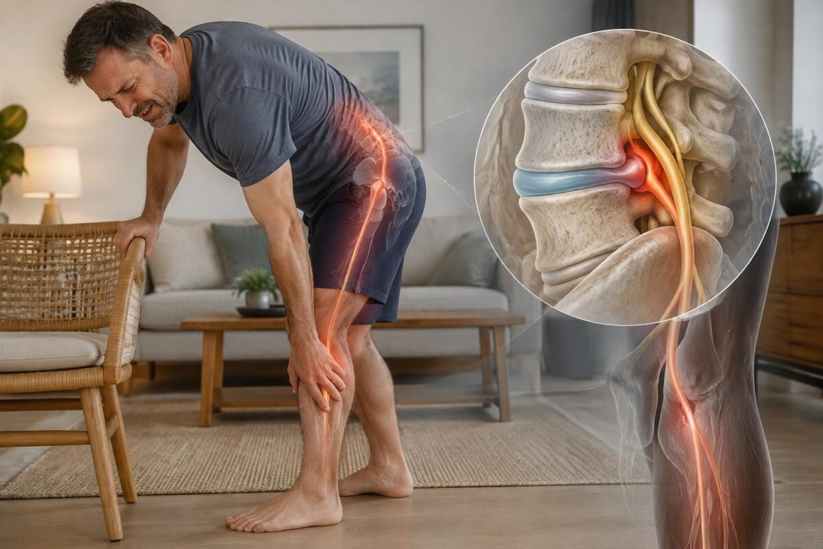
IN BRIEF A disc protrusion is a bulging of a disc that can irritate a nerve root and cause pain radiating down into the buttock and the calf, sometimes with tingling. However, it does not always cause symptoms. Evaluation helps…

Disc herniation and pain behind the thigh
IN BRIEF A disc protrusion is a bulging of a spinal disc, often without symptoms. A pain behind the thigh can be muscular or, if it originates from the back and is accompanied by tingling or numbness, related to nerve…

Disc herniation and pain in the buttock
IN SHORT A disc protrusion is a disc that bulges without its outer layer being ruptured. If it irritates a nerve root, it can contribute to buttock pain, sometimes accompanied by radiation down the leg, tingling, or numbness. It can…

L5-S1 protrusion and sciatic pain: pathway to the foot
IN BRIEF A L5-S1 disc protrusion is a disc that slightly bulges between the lumbar and sacral vertebrae. If it irritates a nerve root, pain may radiate from the lower back or buttock down the back of the leg to…

L4-L5 protrusion and sciatic pain: possible signs
IN BRIEF A L4-L5 disc protrusion is a bulging of the disc between two lumbar vertebrae. If it irritates the L5 nerve root, it can cause sciatica radiating from the buttock to the side of the leg and the top…
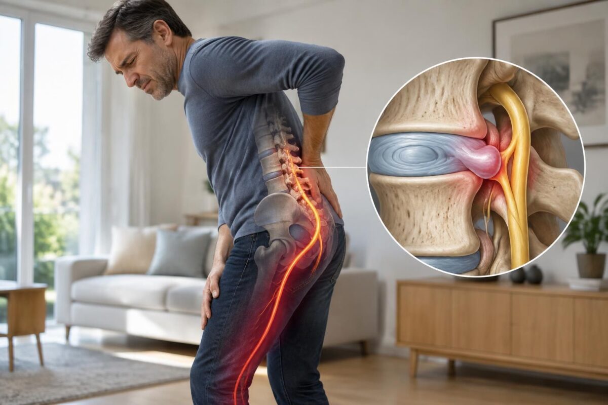
Disc protrusion and sciatica: when the disc touches a nerve root
IN BRIEF A disc protrusion is a disc that extends beyond its usual contour. If it irritates a nerve root, it can contribute to sciatica, with pain radiating into the buttock or leg, sometimes accompanied by tingling or weakness. An…

Dr Sylvain Desforges, B.Sc., D.O., N.D., osteopath, is the founding president of TAGMED and of the ACMA association. He dedicates his practice to chronic spinal pain, low back pain and symptoms compatible with sciatica, particularly when pain radiates toward the buttock, leg, calf, foot or toes.
Through SOS Sciatique, he publishes educational content on sciatica symptoms, possible causes such as disc herniation, disc narrowing, foraminal stenosis or irritation of a lumbar nerve root, as well as non-surgical approaches that may be considered according to the evaluation. At Clinique TAGMED, care may include, depending on the case, motorized neurovertebral decompression, specific osteopathic care or precision percussion.
For an evaluation related to sciatica treatment in Montreal / Mount Royal or sciatica treatment in Terrebonne, please consult the services available at Clinique TAGMED.
Medical disclaimer
The information published on SOS Sciatique is provided for educational purposes only and does not replace a consultation, diagnosis, opinion or treatment provided by a physician or another authorized healthcare professional. The author of this article is neither a medical doctor nor a specialist in a medical specialty recognized by the Collège des médecins du Québec. The osteopathic, manual medicine or functional support approaches described on this site do not constitute medical acts and are not intended to replace medical follow-up.
Consult a physician or emergency service in cases of loss of bladder or bowel control, anesthesia in the saddle area, significant or progressive weakness in the leg or foot, fever, recent trauma, severe pain or rapid worsening of symptoms. For more details, please read our Legal Notice.