


Sciatica and pain during exercise: sign to watch out for
04/11/2026
Sciatica in Terrebonne: why an MRI must be correlated with symptoms
05/11/2026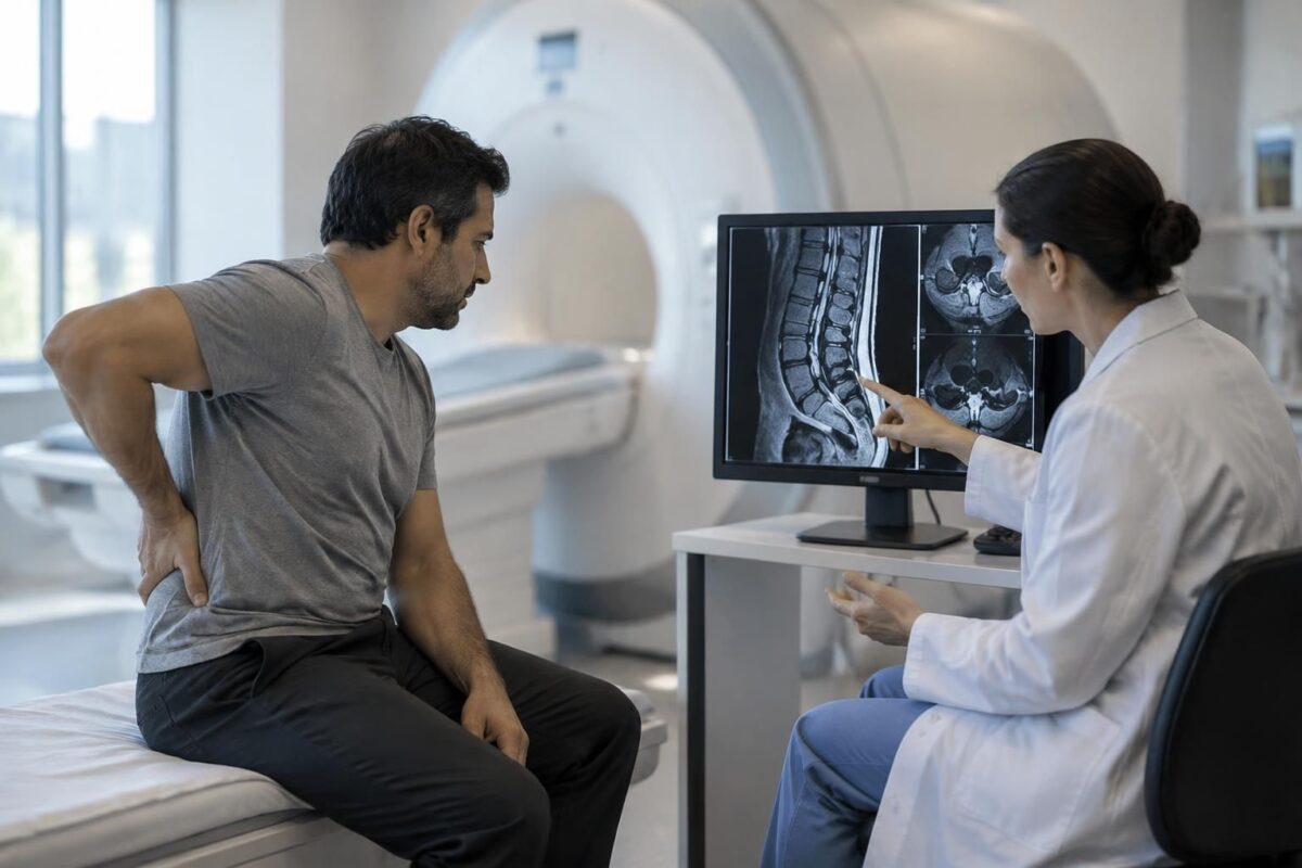
IN BRIEF
| An MRI for sciatica shows structures in the lower back, but an anomaly alone does not prove the cause of the pain. It must be compared with the symptom pathway, the affected side, and the neurological examination. This alignment helps avoid misleading interpretations and choose useful next steps. In Montreal, an evaluation can relate your symptoms to already completed examinations; however, the MRI is not necessary for every episode of sciatica. |
Sciatica in Montreal: why correlate the MRI with symptoms?
An MRI may reveal a disc herniation, a protrusion, or age-related changes without necessarily explaining the pain. To understand their significance, the images must be compared with the symptom pathway, the affected side, and the results of the clinical examination, specifically the strength, sensitivity, and reflexes.
This alignment helps to avoid overestimating a fortuitous anomaly and to identify a neurological deficit requiring prompt attention. At TAGMED Clinic, an evaluation can help examine symptoms and the results already available to discuss appropriate next steps.
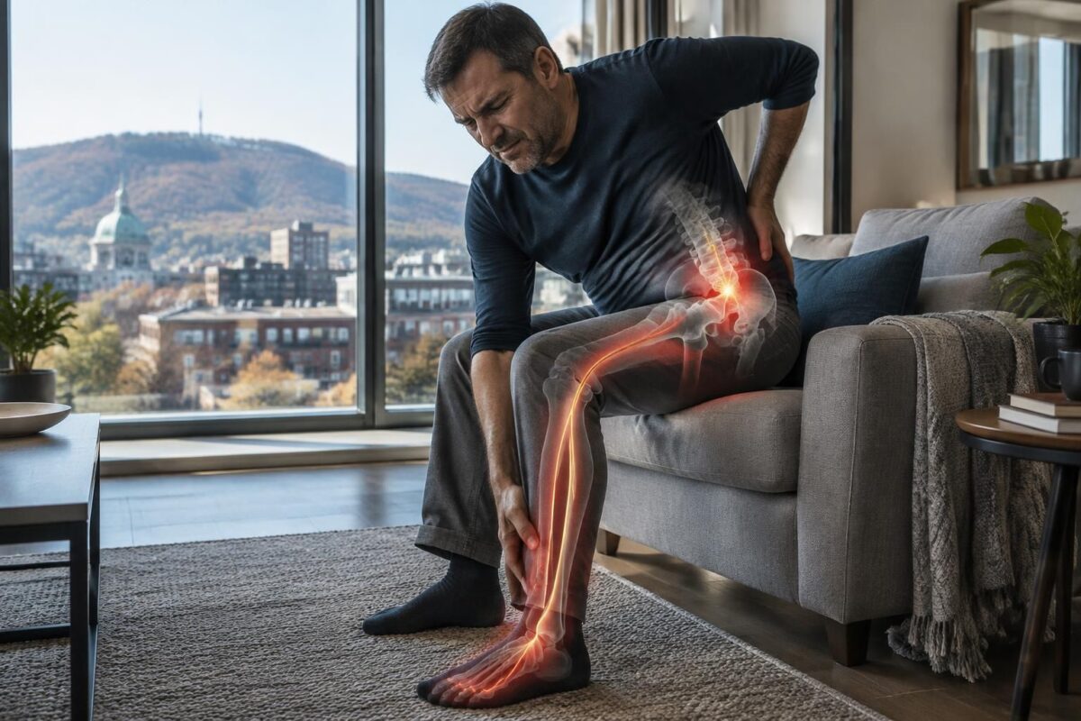
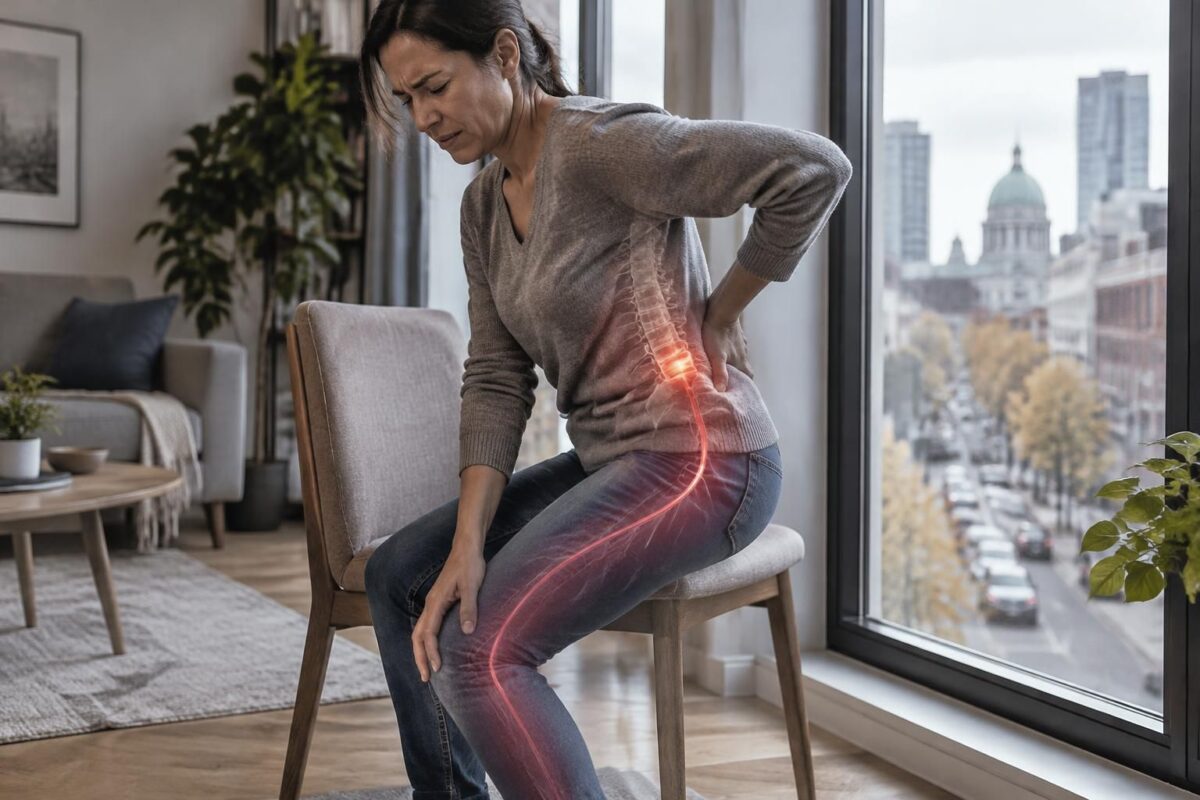
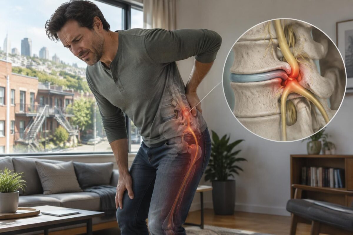
An MRI of the back may reveal a herniation, a disc bulge, or a stenosis, without these results necessarily explaining sciatica. To understand their significance, a professional must compare the images with the pain pathway, the affected side, and the results of the neurological examination. This correlation helps avoid treating a fortuitous anomaly and quickly identify a deficit that requires medical assessment. Sciatica generally refers to pain related to the irritation of a lumbar nerve root. It may radiate from the lower back down to the buttock and leg, sometimes reaching the calf, foot, or toes. Tingling, numbness, or weakness may also occur. The MRI shows structures; it does not directly measure pain and does not confirm, on its own, the cause of the symptoms. An image may show a disc protrusion, a herniation, stenosis, or age-related changes. Some individuals show these results without corresponding pain. Conversely, a person may experience significant pain even if the MRI does not show a clearly responsible anomaly. Therefore, the results must be interpreted in the context of the individual’s history and examination. To better understand these findings, consult our article on disc bulge and sciatica. An anomaly may be a possible cause, but it can also be a finding unrelated to the symptoms. The professional specifies where the pain begins and how far it radiates. They also note the affected side, the duration of symptoms, sensations of numbness or tingling, and activities that change the pain. An anomaly on the left is less easily explained by pain affecting only the right leg. However, the location must be interpreted within the overall picture, as symptoms do not always follow a perfect pattern. Pain that radiates down the leg may sometimes originate from a structure other than the nerve root. To distinguish radicular pain from referred pain, the assessment takes into account the pathway, sensations, and examination results. You can read this explanation about the difference between radicular pain and referred pain. The clinical examination verifies, among other things, the strength of different muscles, skin sensitivity, reflexes, and gait. The professional compares both sides and seeks signs that correspond to a specific nerve root. A weakness in lifting the foot, for example, requires special attention and must be related to the symptoms and imaging. A new or worsening weakness should not be ignored. To better understand the assessment of certain signs, consult the pages on reflexes, strength, and sensitivity and on difficulty lifting the foot. The MRI can locate an anomaly at a specific level, such as L4-L5 or L5-S1. However, the level noted in the report does not always determine, on its own, which root is irritated. The position and type of herniation, as well as nearby structures, influence interpretation. The side and level should correspond as closely as possible to the clinical picture. Lumbar or foraminal stenosis may also reduce the available space near the nerves. The presence of this narrowing does not automatically prove that it is causing the pain. Symptoms, function, and examination remain essential. Consult the information on lumbar stenosis visible on MRI and sciatica, as well as on the relationship between nerve root, disc, and foramen. The MRI provides detailed images of the discs, nerves, and tissues around the spine. It can help when a professional seeks to understand persistent symptoms, assess a suspected significant compression, or prepare for a planned intervention. The choice of examination depends on the clinical question and the likelihood that the result will alter the next steps. An MRI is not necessary for every episode of low back pain or sciatica. In the absence of warning signs or progressive deficits, immediate imaging is generally not the first step. The decision lies with the professional evaluating the situation. It takes into account the evolution of symptoms, care already received, and examination results. A normal MRI does not mean that the pain is imaginary. An abnormal MRI does not prove that the anomaly is the cause of the pain either. The size of a herniation does not predict the intensity of symptoms by itself. To deepen your understanding of the results, consult the page on the correlation between MRI and sciatica symptoms. A prompt medical evaluation is important in the presence of new, significant, or progressive weakness, a dropped foot, or a leg that increasingly gives way. New urinary or bowel problems, loss of sensation in the perineum, significant symptoms on both sides, major trauma, fever, or general deterioration also require urgent attention. In cases of loss of bladder or bowel control, numbness in the saddle region, or rapidly progressive weakness, contact emergency services. These signs should not wait for a routine appointment. To read other guidelines on warning symptoms, consult this article on signs that require rapid evaluation. Bring the report and, if possible, the images from your previous exams. Describe the pain’s path, the affected side, changes in sensitivity, and any observed weakness. It is helpful to specify how long the symptoms have been present and how they influence walking, sleeping, or daily activities. Ask the professional what anomaly could correspond to your symptoms and what elements support this interpretation. You can also inquire if another cause is possible, if the MRI changes the care plan, and what changes should lead to a new evaluation. Follow-up should observe function and neurological signs, rather than being limited to a single image or pain measurement. At the TAGMED Clinic, a consultation can help discuss symptoms, already available exams, and possible next steps. Services depend on the consulted professional, their qualifications, and the evaluated situation. A consultation in osteopathy does not replace a medical diagnosis or an urgent evaluation when a warning sign is present. To learn more about the evaluation and care related to sciatica, check the pages of SOS Sciatica in Montreal or the sciatica treatment in Terrebonne. No. Some hernias are present in people without symptoms. The result becomes more relevant when it corresponds to the side, path of the pain, and signs observed during the examination. Yes. Its position and clinical context may matter more than its size alone. The professional must compare the image with the symptoms and examination. Yes. Size does not perfectly predict pain or loss of function. The evaluation also focuses on localization, neurological signs, and their evolution. The professional may re-examine the hypothesis, consider other causes, or determine if another examination is relevant. Symptoms should not be automatically attributed to an anomaly that does not match the picture. The decision depends on a clinical change and the possibility that the new exam will modify the care. A new MRI is not automatically necessary simply because pain persists. Sylvain Desforges, B.Sc., D.O., N.D., Doctor of Osteopathy and naturopath, is the founding president of TAGMED and the ACMA association. His practice is dedicated to chronic pain of the spine, low back pain, and pain compatible with sciatica, particularly when the pain radiates down to the buttock, leg, calf, foot, or toes. Through SOS Sciatica, he publishes educational content on sciatica symptoms, possible causes such as disc herniation, disc pinching, foraminal stenosis, or irritation of a lumbar nerve root, as well as non-surgical approaches that may be considered based on the evaluation. At the TAGMED Clinic, care may include, depending on the case, motorized neurovertebral decompression, specific osteopathy, or precision percussor. For an evaluation related to the sciatica treatment in Montreal / Mont-Royal or to the sciatica treatment in Terrebonne, check the available services at the TAGMED Clinic. The information published on SOS Sciatica is provided for educational purposes and does not replace a consultation, diagnosis, opinion, or treatment performed by a physician or another licensed healthcare professional. The author of this article is neither a doctor nor a specialist in a medical specialty recognized by the Collège des médecins du Québec. The approaches of osteopathy, manual medicine, or functional support described on this site do not constitute medical acts and are not intended to replace medical follow-up. Consult a physician or an emergency service in case of loss of bladder or bowel control, anesthesia in the saddle area, significant or progressive weakness of the leg or foot, fever, recent trauma, severe pain, or rapid deterioration. For more details, please read our Legal notice. Medical Disclaimer: The information and advice provided on this site do not replace the advice, diagnosis, or treatment of a healthcare professional. Please note that Sylvain Desforges, B.Sc., D.O., N.D., Doctor of Osteopathy, is neither a medical doctor nor a physician, and is not a specialist in a medical specialty as defined by the Collège des médecins du Québec. Manual medicine, functional medicine, and sports medicine as described on this site exclude any medical treatment or diagnosis made by a physician or specialist. Always consult your doctor for any medical questions. For more details, please read our complete Legal Notice.In Montreal, an image is not enough to explain sciatica
How to verify the correlation between the MRI and symptoms
The pathway and side of the pain
Strength, sensitivity, and reflexes
The level observed on the images
What the MRI can provide — and its limits
When rapid investigation or assessment may be necessary
Preparing for a consultation on MRI results
An evaluation in Montreal or Terrebonne
Frequently Asked Questions about MRI and Sciatica
Does a visible hernia on an MRI always cause sciatica?
Can a small hernia cause symptoms?
Can a large hernia cause few symptoms?
What to do if the MRI report and examination do not match?
How often should an MRI be repeated?
About the Author
Medical Disclaimer
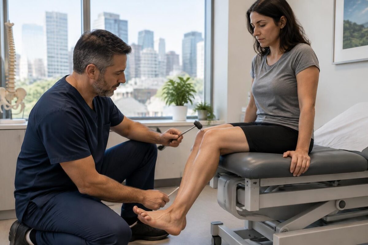
Sciatica in Montreal: reflexes, strength, and sensitivity in evaluation
IN BRIEF The evaluation of sciatica involves examining reflexes, muscle strength, and sensitivity, in addition to the pathway and evolution of pain. In Montreal, these observations help the healthcare professional identify potential neurological signs and guide the next steps. No…
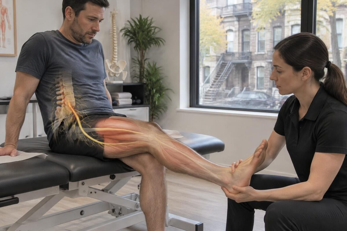
Sciatica in Montreal: Muscle Weakness Based on the Irritated Root
IN BRIEF Sciatica can cause muscle weakness when lumbar nerve roots are irritated. Depending on the affected root, it may become difficult to lift the foot or stand on tiptoes. In Montreal, an assessment of strength, sensation, and pain pathway…
Sciatica in Montreal: why correlate MRI with symptoms?
Shows structures, not always the cause of pain.
The path, side, and numbness guide the evaluation.
Strength, sensitivity, reflexes, and walking complete the analysis.
An anomaly is more relevant if it corresponds to the observed signs.
A hernia or wear on the MRI may be present without symptoms.
Imaging should inform a care decision, not replace it.

In Montreal, a lumbar MRI can reveal a disc herniation, a protrusion, or stenosis, but these images alone do not prove the cause of sciatica. To guide the next steps, the results must match the pain pathway, the affected side, and the signs noted during the clinical examination. A comprehensive assessment helps avoid neglecting a neurological deficit as well as attributing symptoms to an anomaly with no certain link.
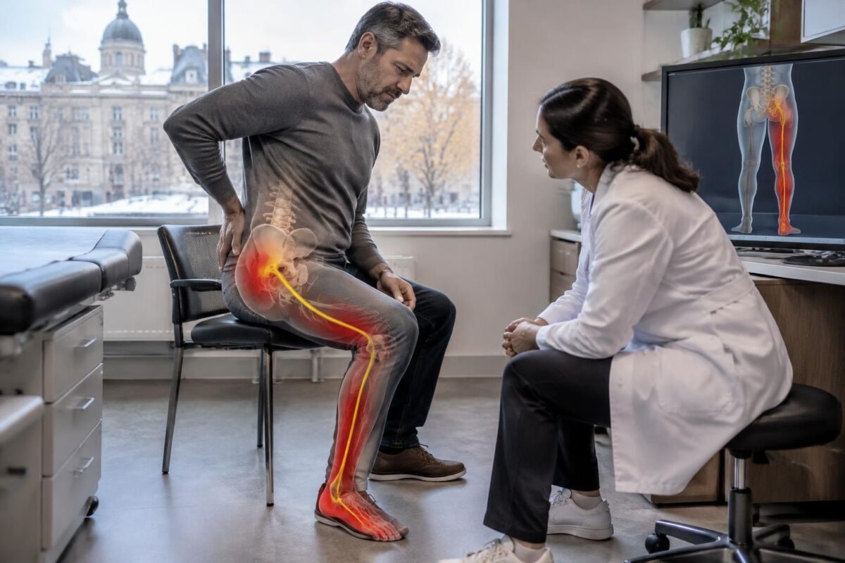
Sciatica in Montreal: nerve pain and sensory territory
IN BRIEF Sciatica is a nerve pain that radiates from the lower back or buttock down the leg. Its sensitive territory varies depending on the affected root and may include burning, tingling, or numbness down to the foot. In Montreal,…
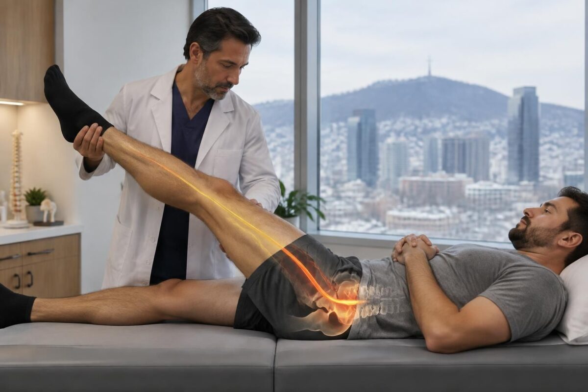
Sciatica in Montreal: irritation L5 or S1, why the clinical examination matters
IN BRIEF The sciatica related to irritation of the L5 or S1 root can cause pain, numbness, or weakness in the leg and foot. In Montreal, the clinical examination compares the course of symptoms, sensitivity, strength, and reflexes to guide…
Why an anomaly on the MRI is not enough
Imaging studies can show disc or joint changes in people who feel no pain. Some of these changes also become more frequent with age. Thus, the presence of a hernia on the MRI does not automatically mean it explains lower back pain or pain that radiates to the buttock and leg.
The interpretation must consider several elements: the location of the anomaly, the affected side, the reported symptoms, and the results of the physical examination. A finding is more relevant when these clues are consistent with each other and can genuinely influence care.
Sciatica in Montreal: L5-S1 Herniation and Pain Towards the Foot
IN BRIEF A L5-S1 disc herniation can irritate the S1 nerve root and cause sciatica in Montreal, with pain or numbness radiating down to the calf, heel, or foot. The assessment correlates symptoms, strength, and sensitivity with imaging, if relevant;…
Sciatica in Montreal: L4-L5 protrusion and leg pain
IN BRIEF An L4-L5 disc protrusion is a bulging of the disc that can irritate a nerve root and cause sciatica, with pain in the buttock and leg. However, it does not always cause symptoms. In Montreal, an assessment can…
Elements to compare with the MRI
The pathway and nature of symptoms
Describe precisely where the pain starts and how far it descends: buttock, thigh, calf, foot, or toes. Specify also the duration of symptoms, their intensity, and the positions or activities that worsen or relieve them. Numbness, tingling, and any sensation of weakness are also important to report.
The neurological examination and function
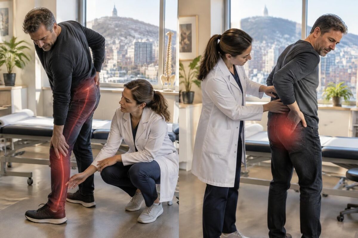
A qualified professional can examine strength, sensitivity, reflexes, and gait, comparing both sides. These observations help verify if the signs are consistent with irritation of a nerve root and if function is changing. The side and level described in the MRI report must be correlated with these findings, rather than considered separately.
Sciatica in Montreal: what “disc-root conflict” means
IN BRIEF A disc-root conflict refers to possible contact or pressure between an intervertebral disc and a nerve root. In Montreal, this mention in the MRI can help understand a sciatica, but does not by itself prove that the disc…
Sciatica in Montreal: radicular pain versus referred pain
IN BRIEF Radicular sciatica corresponds to irritation of a nerve root, while referred pain can spread to the buttock or leg without direct nerve involvement. In Montreal, distinguishing these pains helps guide the evaluation by linking the symptom route to…
When can the MRI be useful?
Imaging is not necessarily the first step during a recent and stable episode, in the absence of warning signs. It becomes more relevant if symptoms persist despite appropriate management, if a neurological deficit is observed, or if the result can change the care plan, especially before considering an intervention. A normal MRI does not negate the pain; conversely, an abnormal image does not confirm its origin alone.
Before the examination, ask yourself with the professional who is following you: what question should the MRI clarify? What result could change the proposed course? A new MRI is generally not decided solely because a period has elapsed: the evolution of symptoms and the possibility of modifying care matter more.
Monitoring progress and choosing the next step
To assess progress, note changes in strength and sensitivity, the tolerated walking distance, sleep, and daily activities. Bring your old reports and, if possible, your images to appointments so they can be compared to the current clinical picture. An assessment at the TAGMED Clinic in Montreal / Mont-Royal can help relate symptoms, function, and available exams, then clarify appropriate options. The services offered depend on each situation and the skills of the concerned professional.
When to seek help without delay
Quickly consult a doctor or emergency service in case of new, significant, or progressive weakness, foot drop, new urinary or bowel problems, or numbness in the perineal area. Rapid deterioration, major trauma, fever, or a general state that worsens also necessitate urgent evaluation; these signs should not wait for a routine appointment.
Medical warning: This content is educational and does not replace a consultation, diagnosis, or treatment by a licensed healthcare professional. The author is neither a doctor nor a specialist in a medical specialty recognized by the Collège des médecins du Québec. Approaches in osteopathy or functional support do not replace medical follow-up.
| Clinical marker | Why compare it to the MRI |
|---|---|
| Pain pathway | Verify that the affected side and area match. |
| Numbness or tingling | Compare their distribution to the observed level. |
| Strength of the leg and foot | Identify a deficit compatible with nerve compression. |
| Reflexes and sensitivity | Confirm if the exam results support the hypothesis. |
| Hernia or disc protrusion | An anomaly on the image does not prove it causes the pain. |
| Stenosis or degenerative changes | Relate them to the symptoms and impaired function. |
| Normal MRI without explanatory anomaly | The pain can be real; other causes must be considered. |
| New or progressive weakness | A rapid medical evaluation may be necessary. |
What the image shows is not enough to explain the pain
The testimonies below are fictional examples for educational purposes: they illustrate possible situations and do not correspond to real patients. They do not allow for a diagnosis. For sciatica in Montreal or Mont-Royal, the interpretation of an MRI benefits from considering the pain pathway, the clinical examination, and the evolution of symptoms.
“My MRI shows a hernia, but the pain is on the other side.” In this fictional scenario, the person discovers a disc anomaly in their report, but their symptoms do not seem to match the described side. A hernia or protrusion may be present without causing pain. The professional must therefore verify if the observed level and side are consistent with the symptoms and clinical signs before attributing a role to them.
“I was told I have a large hernia: is it necessarily serious?” Not necessarily. The size of a hernia does not solely predict the intensity of pain or limitations. Its location, the structures involved, muscle strength, sensitivity, and leg function all contribute to the interpretation. An important finding in the image must be evaluated in context, without minimizing symptoms or concluding too quickly.
“My MRI shows changes, but I don’t have pain radiating down my leg.” The images may reveal signs of wear, a disc bulge, or stenosis without necessarily explaining the current symptoms. The clinical examination helps distinguish a common anomaly from a problem possibly related to a pain pathway, numbness, or weakness.
“The exam and the MRI do not tell the same story.” A discrepancy is a reason to reassess the situation, not to force an explanation. It may be necessary to review the history, compare both sides, consider other causes, or discuss the relevance of additional examinations. The MRI describes the anatomy; it does not confirm on its own the exact source of every pain.
How to link symptoms, the exam, and imaging?
The evaluation begins with a precise description of the symptoms: their path — lower back, buttock, thigh, calf, or foot —, their duration, intensity, and the factors that aggravate or relieve them. Tingling, areas of numbness, and any weakness should also be mentioned.
The clinical exam can compare strength, sensitivity, reflexes, and walking between the sides, then use targeted tests according to the observed findings. If an MRI or CT scan is available, its findings are related to the affected side, the concerned level, and the functions that are actually disrupted. An electromyogram may sometimes be considered when additional functional information is useful or when the differential diagnosis remains uncertain.
Before requesting or repeating an MRI, the useful question is whether its result could change the course of action. For many recent and stable episodes, immediate imaging is not automatically necessary. It may become more relevant if symptoms persist despite care, if a neurological deficit is observed, or if an intervention is being considered. The choice and timing of the examination depend on an evaluation by a licensed professional.
Follow-up should not rely on a single image or a single pain rating. It can also take into account the evolution of strength, sensitivity, walking, sleep, tolerance to sitting, and important daily activities. Bringing old reports or images can facilitate comparison.
When to consult quickly?
A new, significant, or rapidly progressive weakness, a dropped foot, a leg that increasingly gives way, significant symptoms on both sides, or rapid deterioration require a prompt evaluation. New urinary or intestinal issues, or numbness in the saddle region or perineum, require urgent consultation. Fever, major trauma, or a general state of deterioration also justify not waiting for a routine appointment.
At the TAGMED Clinic, an evaluation related to sciatica can help put symptoms, exam findings, and previous examinations into perspective to discuss appropriate next steps. The care provided depends on each situation and the qualifications of the concerned professional; it does not replace a diagnosis or medical follow-up.
About the author
Sylvain Desforges, B.Sc., D.O., N.D., Doctor of Osteopathy and naturopath, is the founding president of TAGMED and the ACMA association. His practice focuses particularly on chronic spinal pain, low back pain, and symptoms consistent with sciatica.
Medical disclaimer
This content is provided for educational purposes and does not replace consultation, diagnosis, advice, or treatment by a physician or other licensed health professional. The author is neither a doctor nor a specialist in a medical specialty recognized by the Collège des médecins du Québec. Approaches involving osteopathy, manual medicine, or functional support are not medical acts and do not replace medical follow-up.
When low back pain radiates to the buttock, leg, or down to the foot, an MRI can help examine the structures of the spine. It may reveal a herniated or bulging disc, stenosis of the canal, or nerve openings, as well as changes associated with aging. However, the imaging report alone is not sufficient to explain sciatica.
Visible anomalies on an MRI may sometimes be present in people who feel no pain. Conversely, a person may experience real symptoms even if the imaging does not show an obvious cause. This is why each result must be interpreted in light of the symptom history, their evolution, and the physical examination. The image provides anatomical information; it does not directly measure pain or its consequences on daily activities.
Clinical concordance is particularly important. The professional examines whether the side and level of the anomaly correspond to the pain path, areas of numbness or tingling, and any weaknesses. They may also compare strength, sensitivity, and reflexes on both sides, observe walking, and perform targeted tests. An anomaly consistent with multiple clinical findings is generally more relevant than an isolated finding.
The size of a hernia does not, on its own, predict the intensity of the symptoms. A small hernia can be painful depending on its position and the structures it irritates; a larger hernia can sometimes be minimally symptomatic. Similarly, a vertebral level indicated in the report does not automatically designate a specific nerve root: the location and shape of the lesion also matter. If the MRI and the exam do not tell the same story, the working diagnosis may need to be reevaluated.
An MRI is not necessarily the first examination for every episode of low back pain or sciatica. When there are no red flags and the symptoms are recent or stable, immediate imaging may not change the management. It becomes more relevant if symptoms persist despite care, if a neurological deficit is observed, or if an intervention is being considered. Therefore, the useful question is: could the result change the next steps? An additional examination, such as an electromyogram, may sometimes be discussed when functional information is needed or when the diagnosis remains uncertain.
To prepare for an evaluation in Montreal, accurately describe the pain path, duration, aggravating or relieving factors, numbness, and changes in strength. Bring the report and any previous images, if you have them. Follow-up can then focus on measurable elements: strength, sensitivity, walking, sleep, and ability to sit or perform important activities.
Consult quickly or urgently in the presence of significant or progressive weakness, a new dropped foot, urinary or intestinal issues, or numbness in the saddle region. Rapid deterioration, major trauma, fever, or a general state of worsening also requires evaluation without waiting for a routine appointment.
About the author
Sylvain Desforges, B.Sc., D.O., N.D., Doctor of Osteopathy and Naturopath, is the founding president of TAGMED clinics and the ACMA association. His practice particularly focuses on chronic spinal pain and symptoms consistent with sciatica. His approach emphasizes assessment, function, and adapting care to each person’s situation. Depending on the case and the professionals involved, services may include specific osteopathy or motorized neurovertebral decompression.
Medical Disclaimer
This content is provided for educational purposes and does not replace a consultation, diagnosis, or treatment by a physician or another authorized healthcare professional. The author is neither a doctor nor a specialist in a medical specialty recognized by the Collège des médecins du Québec. The approaches described do not constitute medical acts and do not substitute for medical follow-up.
In Montreal, MRI can help identify a herniated disc, stenosis, or other lumbar changes, but these images alone do not explain sciatica. Their value depends on their alignment with the pain path, neurological signs, and clinical examination results.
A lumbar MRI is an anatomical photograph, not automatic proof of the cause of pain. Disc protrusions, osteoarthritis, or signs of wear can appear even in people who have no corresponding symptoms. To interpret a result, it is necessary to verify whether the observed anomaly corresponds to the affected side, the relevant lumbar level, and the actual difficulties present. The assessment of an abnormal lumbar MRI illustrates why images must be placed back into their clinical context.
This connection starts with a precise description of the symptoms: where the pain starts, how far it travels down, whether it is accompanied by tingling or numbness, and what movements or positions alter it. Pain heightened in a sitting position may guide the evaluation toward certain hypotheses, though it is not sufficient to establish a diagnosis; see also sciatica aggravated in a sitting position. Tolerance to exercise and the distribution of symptoms in the leg are also factors to examine, as explained in this article about sciatic pain during exercise.
The clinical examination completes this story by assessing notably strength, sensitivity, reflexes, and gait. An observation on the MRI becomes more convincing when several elements tell the same story: for example, an anomaly located on the correct side and at a level compatible with the painful area and the observed signs. However, the relationship between a disc level and a nerve root is not always mechanical; L5-S1 and sciatica as well as information about sensitive areas associated with nerve pain can help understand why the path of symptoms matters so much.
An MRI is not necessarily the first step for every episode of low back pain or sciatica. It is particularly useful when symptoms persist despite appropriate management, when a neurological sign requires further investigation, or when the result could change treatment options. Resources on the circumstances when an MRI may be relevant for back pain and what a lumbar MRI can reveal in the presence of symptoms in the leg provide additional benchmarks. If the MRI and the examination do not match, it is prudent to reassess the initial hypothesis rather than automatically attributing the pain to a visible anomaly. Depending on the clinical question, a professional may also determine if another test, such as an EMG, is appropriate.
Follow-up should focus on concrete changes: strength, sensitivity, gait, sleep, and ability to resume important activities. Generic exercises may not be suitable for all presentations; the choice must consider symptoms and function, as highlighted by this resource on exercises for sciatica. To better connect symptoms, examination, and already performed tests, a sciatica assessment in Montreal can help clarify the next steps. A discussion on the MRI of the sciatic nerve and its indications is also offered by this imaging resource.
Finally, a new or rapidly progressing weakness, a dropped foot, loss of bladder or bowel control, or numbness in the perineal area requires urgent medical evaluation. In the absence of these signs, the goal remains to choose tests and care based on the overall picture, rather than an isolated image. An individualized interpretation helps avoid both trivializing a neurological deficit and overinterpreting a chance anomaly.

Sylvain Desforges, B.Sc., D.O., N.D., docteur en ostéopathie
Dr Sylvain Desforges, B.Sc., D.O., N.D., osteopath, is the founding president of TAGMED and of the ACMA association. He dedicates his practice to chronic spinal pain, low back pain and symptoms compatible with sciatica, particularly when pain radiates toward the buttock, leg, calf, foot or toes.
Through SOS Sciatique, he publishes educational content on sciatica symptoms, possible causes such as disc herniation, disc narrowing, foraminal stenosis or irritation of a lumbar nerve root, as well as non-surgical approaches that may be considered according to the evaluation. At Clinique TAGMED, care may include, depending on the case, motorized neurovertebral decompression, specific osteopathic care or precision percussion.
For an evaluation related to sciatica treatment in Montreal / Mount Royal or sciatica treatment in Terrebonne, please consult the services available at Clinique TAGMED.
Medical disclaimer
The information published on SOS Sciatique is provided for educational purposes only and does not replace a consultation, diagnosis, opinion or treatment provided by a physician or another authorized healthcare professional. The author of this article is neither a medical doctor nor a specialist in a medical specialty recognized by the Collège des médecins du Québec. The osteopathic, manual medicine or functional support approaches described on this site do not constitute medical acts and are not intended to replace medical follow-up.
Consult a physician or emergency service in cases of loss of bladder or bowel control, anesthesia in the saddle area, significant or progressive weakness in the leg or foot, fever, recent trauma, severe pain or rapid worsening of symptoms. For more details, please read our Legal Notice.