

Sciatica in Montreal: why an MRI must be correlated with symptoms
05/11/2026
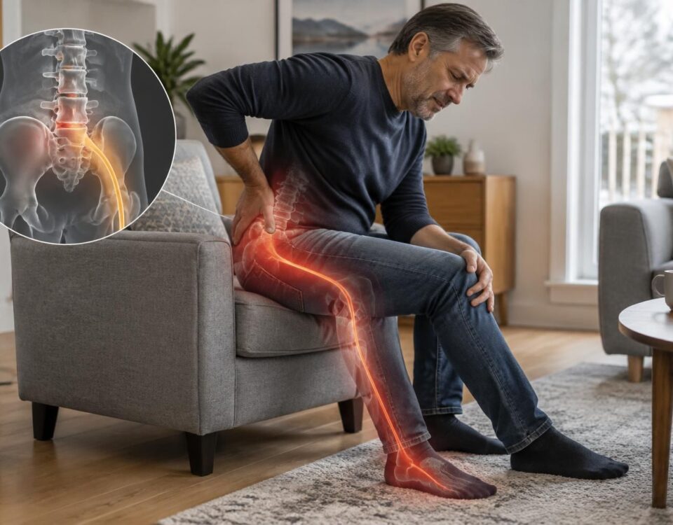
Acute sciatica: typical symptoms to recognize
05/11/2026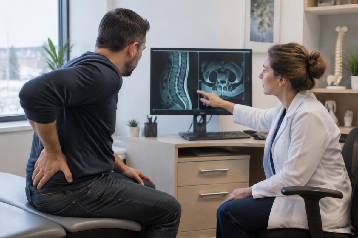
IN BRIEF
| Sciatica is a pain that often radiates from the lower back to the buttock and leg. A lumbar MRI shows the structures of the spine, but its results must correspond to the symptoms and clinical examination: a visible anomaly does not always explain the pain. In Terrebonne, this correlation helps guide management and determine whether further examinations or treatments are indicated. |
Sciatica in Terrebonne: Why Correlate MRI With Symptoms
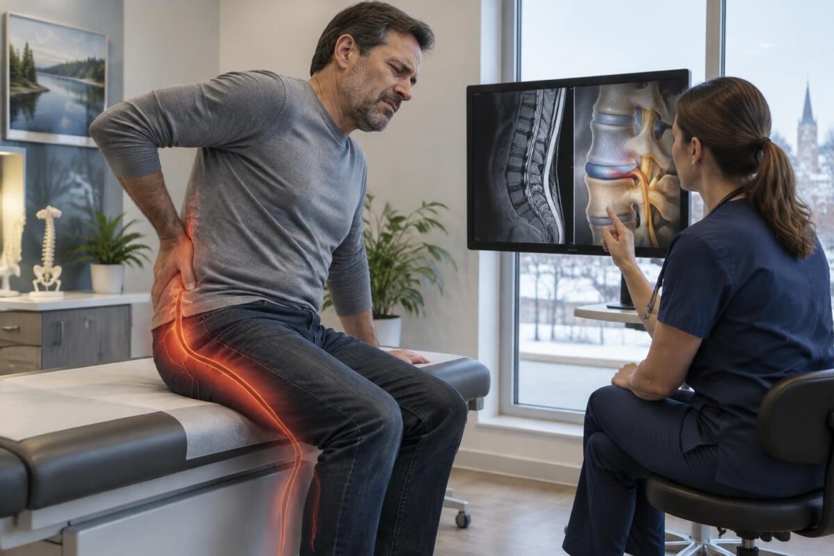
A lumbar MRI can show a herniated disc or other changes that are not necessarily the cause of the pain. To interpret the image correctly, a healthcare professional compares it to the symptoms, their path in the leg, and the results of the clinical examination. This correlation helps prevent attributing sciatica to an incidental finding and guides management.
An MRI is not routinely performed at the onset of sciatica; its usefulness depends on the progression, observed signs, and envisaged treatments. Progressive weakness, loss of bladder or bowel control, or numbness in the saddle area requires urgent medical evaluation.
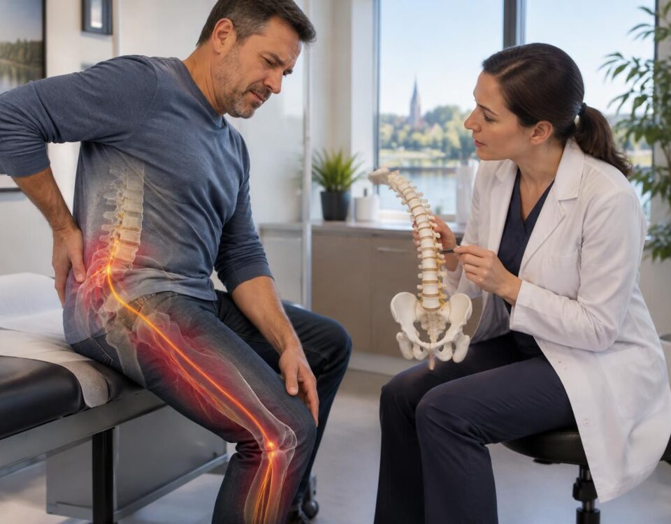
An MRI can show a disc bulge, a herniation, or a narrowing, but these images do not always prove the cause of sciatica. To interpret the result, a healthcare professional must compare the images to the path of the pain, changes in sensitivity, and muscle strength. This concordance helps choose the next steps and identify situations requiring rapid evaluation. A lumbar MRI produces detailed images of the discs, vertebrae, and structures near the nerves. It can reveal a herniated disc, a bulge, disc degeneration, or narrowing of the space where a nerve root passes. These changes do not always cause symptoms. Some individuals may show an anomaly on an MRI without experiencing pain. Conversely, significant pain may necessitate evaluation even if the image does not show obvious compression. An imaging result must therefore be interpreted alongside the clinical examination and the history of symptoms. A herniation may occur at a different level from where the pain travels. An anomaly may also be present without irritating a nerve root. The professional checks whether the level and side shown on the MRI correspond to the observed signs. This comparison is sometimes referred to as the concordance between MRI and symptoms. To learn more about this approach, consult the TAGMED Clinic page on the concordance between MRI and symptoms. Sciatica generally corresponds to pain that starts from the lower back or buttock and descends into part of the leg. It may be accompanied by tingling, numbness, or weakness. The exact location of symptoms provides clues about the nerve root that may be irritated, without confirming the diagnosis on its own. Pain in the buttock can sometimes be related to nerve irritation, even if the lower back does not hurt. Other causes may also explain this symptom. The professional must consider the entire path of the pain and other signs before linking the pain to an anomaly on the MRI. Consult this article on pain in the buttock without back pain. Pain that descends towards the top of the foot, heel, or toes can help specify the affected area. However, sensation alone does not allow for the certain identification of the level of the spine involved. Examination of strength, sensitivity, and reflexes completes the analysis. To better understand these paths, consult the pages on pain toward the top of the foot associated with the L4-L5 level and on persistent pain in the heel. A sharp pain or electric shock sensation in the calf may suggest nerve-type pain. This sign must be evaluated with other symptoms. It does not, on its own, conclude that a herniation visible on the MRI is responsible. For more details, read the page on electric shock sensation in the calf. Lumbar nerve roots transmit sensations and motor signals to different parts of the leg and foot. Irritation at a given level can therefore produce symptoms in a zone distant from the lower back. The professional compares the side and level of the anomaly on the MRI with the distribution of symptoms and the results of the examination. The L4-L5 and L5-S1 levels are often examined when a person presents with low back pain radiating into the leg. However, an image showing a herniation at one of these levels is not sufficient to establish a link with the symptoms. To understand lumbar levels on an MRI, consult this page on reading lumbar levels. An MRI is not necessary for every recent episode of sciatica. The professional usually begins by collecting the history and examining the person. They specifically check the path of pain, sensitivity, strength, reflexes, and the ability to walk. An MRI may be considered if symptoms persist, worsen, or do not improve with initial management. It may also be requested if a neurological deficit is present or if an injection or surgery is considered. The timing of the examination depends on the clinical picture and the opinion of the professional following the individual. Imaging can reveal changes that do not cause pain. It is therefore important to avoid drawing conclusions from an isolated report. This article also explains when an MRI may be useful and what signs should alert. Certain symptoms require urgent medical evaluation. Quickly consult a doctor or an emergency service in cases of significant or progressive weakness in a leg or foot, new difficulty walking, loss of bladder or bowel control, or numbness in the saddle area. Severe pain that worsens, fever, or pain occurring after recent trauma also requires rapid medical attention. In these situations, do not wait for a routine consultation to have the symptoms evaluated. An evaluation begins by listening to symptoms and examining mobility, sensitivity, and strength. The professional may then review the report and MRI images to check if the results match the clinical picture. The decision to request or repeat imaging is up to the healthcare professional authorized to assess the situation. At the Clinique TAGMED, the services offered may include, depending on the evaluation and the person’s situation, specific osteopathy, motorized neurovertebral decompression, or the precision hammer. These approaches do not replace a medical diagnosis and are not suitable for all cases. Signs of severity must first be evaluated by a doctor or an emergency service. To learn about the services related to the treatment of sciatica in Terrebonne, consult the available resources and request an evaluation tailored to your situation. Sylvain Desforges, B.Sc., D.O., N.D., Doctor of Osteopathy and Naturopath, is the founding president of TAGMED and the ACMA association. He dedicates his practice to chronic pain of the spine, lower back pain, and pain consistent with sciatica, particularly when the pain radiates towards the buttock, leg, calf, foot, or toes. Through SOS Sciatique, he publishes educational content about the symptoms of sciatica, possible causes such as herniated discs, disc pinching, foraminal stenosis, or irritation of a lumbar nerve root, as well as non-surgical approaches that may be considered based on the evaluation. At Clinique TAGMED, care may include, depending on the case, motorized neurovertebral decompression, specific osteopathy, or the precision hammer. For an evaluation related to the treatment of sciatica in Montreal / Mont-Royal or the treatment of sciatica in Terrebonne, check the available services at the Clinique TAGMED. Medical disclaimer: The information and advice provided on this site do not replace the opinion, diagnosis, or treatment of a healthcare professional. Please note that Sylvain Desforges, B.Sc., D.O., N.D., Doctor of osteopathy, is neither a doctor of medicine nor a physician, and is not specialized in a medical specialty as defined by the Collège des médecins du Québec. Manual medicine, functional medicine, and sports medicine as described on this site exclude any treatment or medical diagnosis made by a doctor or specialist physician. Always consult your doctor for any medical questions. For more details, please read our Full Legal Notice. The information published on SOS Sciatique is provided for educational purposes and does not replace a consultation, diagnosis, opinion, or treatment performed by a doctor or another authorized healthcare professional. The author of this article is neither a doctor nor a specialist in a medical specialty recognized by the Collège des médecins du Québec. The approaches of osteopathy, manual medicine, or functional support described on this site do not constitute medical acts and are not intended to replace medical follow-up. Consult a doctor or emergency service in the event of loss of bladder or bowel control, numbness in the saddle area, significant or progressive weakness of the leg or foot, fever, recent trauma, severe pain, or rapid worsening. For more details, please read our Legal Notice.What the MRI Can Show
An Abnormal Image Is Not Sufficient to Establish the Cause
The Path of Pain Helps Interpret Results
Pain in the Buttock Without Back Pain
Pain Towards the Top of the Foot or Heel
Electric Shock Sensation in the Calf
Why Lumbar Level Matters
When Is an MRI Useful?
Signs Requiring Rapid Evaluation
How the evaluation is conducted in Terrebonne
About the Author
Medical Disclaimer
Sciatica in Terrebonne: correlate the MRI with the symptoms
An image is interpreted alongside the clinical exam: a visible anomaly on the MRI does not prove, on its own, that it is the cause of the pain.
- What the MRI shows
- What the evaluation verifies
- A herniated disc and its level
- If the level corresponds to the path of pain in the buttock and leg
- A contact or nerve compression
- The presence of tingling, numbness, or concordant weakness
- Occasionally common disc changes
- If they truly explain the symptoms or are an unrelated finding
- An image to be interpreted according to the context
- The clinical exam, evolution, and neurological signs
In case of progressive weakness, loss of bladder or bowel control, or numbness in the saddle area, seek medical evaluation quickly.

In Terrebonne, a lumbar MRI may reveal a herniated disc or another change in the spine, but the image alone is not enough to explain sciatica. To guide treatment, the healthcare professional must compare the examination results with the symptoms, their path, and the clinical exam. This correlation helps avoid misattributing pain to an anomaly that is not linked to it.
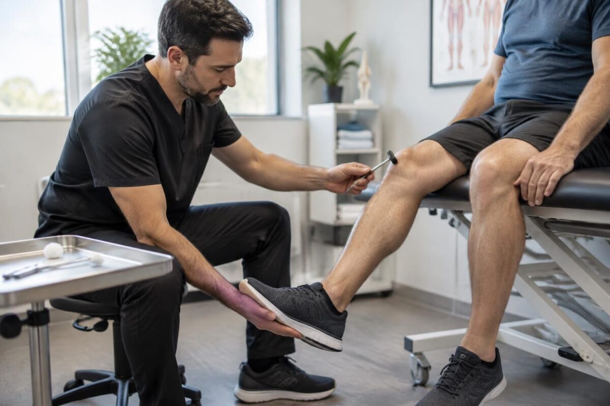
Sciatica in Terrebonne: reflexes, strength, and sensitivity in evaluation
IN BRIEF In Terrebonne, the assessment of sciatica checks the reflexes, strength, and sensitivity of the legs to identify possible nerve damage. These observations, combined with the pain’s pathway and its evolution, help to guide the next steps and recognize…
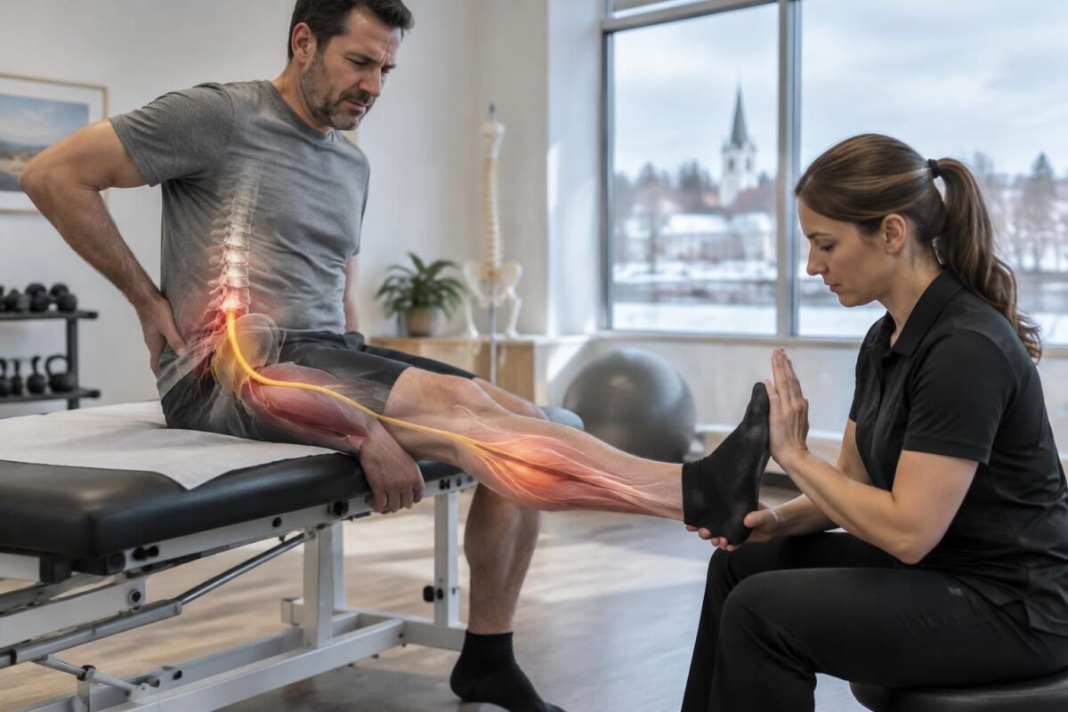
Sciatica in Terrebonne: muscle weakness according to the irritated nerve root
IN BRIEF Sciatica with muscle weakness can occur when a lumbar nerve root is irritated; affected movements, such as raising the foot or walking on tiptoes, provide clues but are not sufficient for a diagnosis. In Terrebonne, an evaluation can…
An anomaly on the MRI is not always the cause of the pain
The MRI shows the discs, nerve roots, and surrounding tissues. It may highlight a hernia, a disc bulge, or signs of wear. However, some people have such anomalies without experiencing pain. Conversely, the intensity of the pain does not always correspond to the extent of what appears on the images.
An imaging result must therefore be interpreted with caution. For example, a hernia visible at a given level is more likely to explain sciatica if its position corresponds to a nerve root and the path of the symptoms. The presence of a hernia alone does not confirm that it is responsible for the pain.
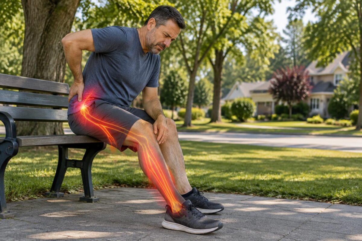
Sciatica in Terrebonne: nerve pain and sensory territory
IN BRIEF Sciatica is a nerve pain that can start from the lower back and radiate into the buttock and leg, sometimes down to the foot, with tingling or numbness. In Terrebonne, identifying the affected sensory territory helps guide the…
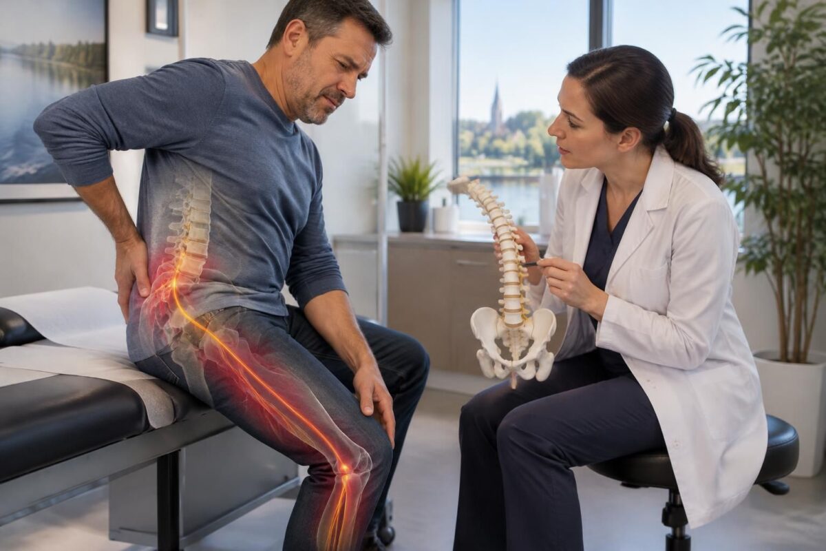
Sciatica in Terrebonne: L4, L5, S1, how to find your way?
IN SHORT Sciatica is a pain that can radiate from the buttock down the leg when a nerve root is irritated. The levels L4, L5, and S1 can produce overlapping symptoms; therefore, the pathway of the pain alone is not…
Compare the image to the path of the symptoms
The territory of pain
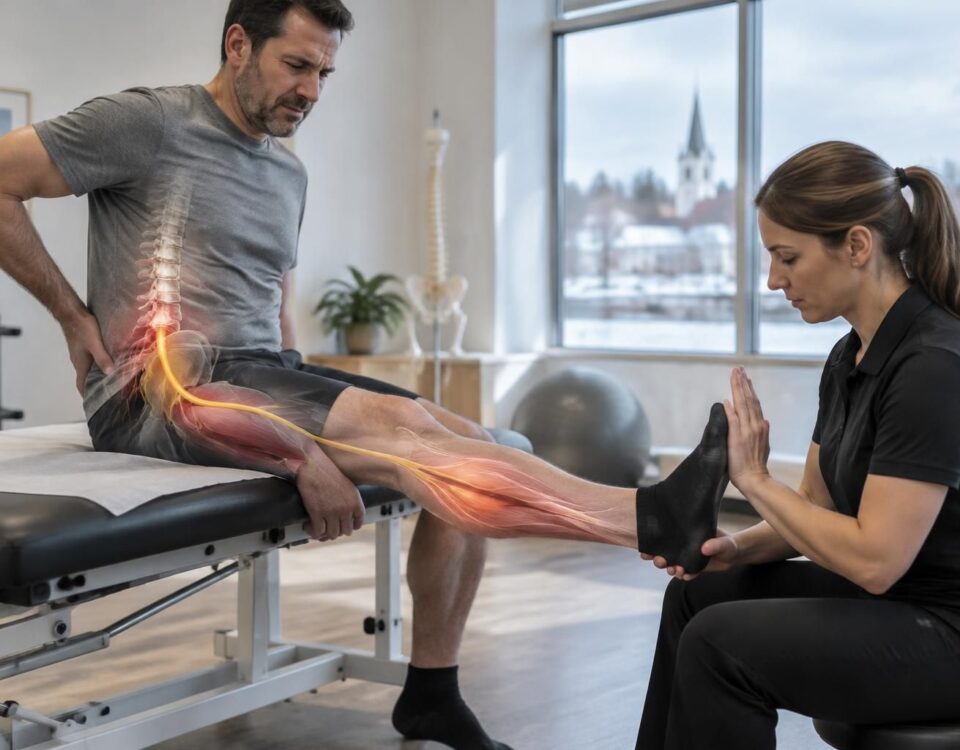
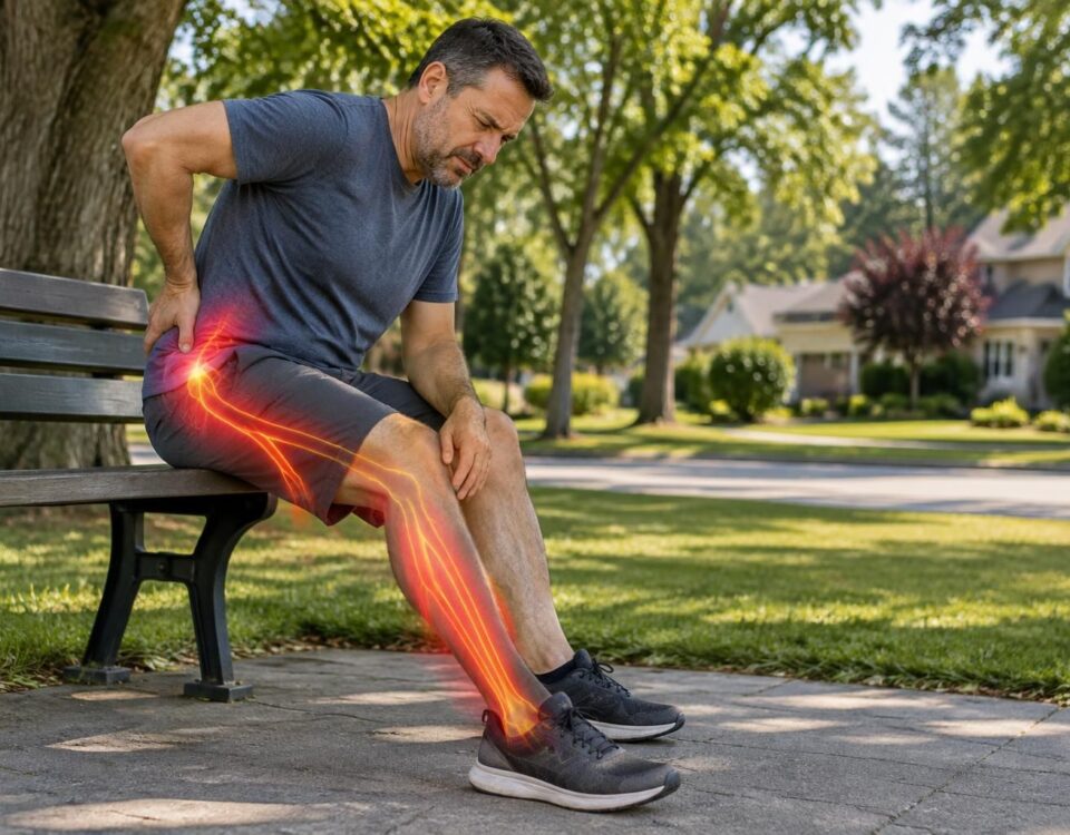
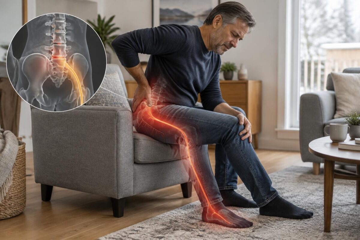
Sciatica generally manifests as pain starting from the lower back or buttock and descending into the leg. It may be accompanied by tingling, numbness, or a feeling of weakness. The pathway varies depending on the irritated nerve root: the doctor checks if the symptoms match the level and side of the anomaly observed on the MRI.
Location also matters. An image showing an issue on the right side does not automatically explain symptoms primarily on the left. Similarly, an anomaly located at a level that does not correspond to the painful pathway may be an incidental finding. Examination of strength, sensitivity, reflexes, and gait provides essential complementary information.

Sciatica in Terrebonne: L5 or S1 irritation, why the clinical examination matters
IN BRIEF Sciatica is pain related to the irritation of a nerve root, often L5 or S1. The pathway of symptoms, strength, sensitivity, and walking provide clues, but the territories may overlap: the clinical examination therefore helps guide the assessment…
Sciatica in Terrebonne: L5-S1 Herniation and Pain Towards the Foot
IN BRIEF A sciatica related to a possible L5-S1 herniation can cause pain from the lower back or buttock down to the calf, heel, or foot. The path of the symptoms alone is not enough to confirm the cause: the…
When is an MRI relevant?
In the absence of concerning signs, an MRI is not always necessary in the first days of sciatica. The professional may first assess the symptoms and propose appropriate management, then monitor their evolution. Imaging can be considered if pain persists, worsens, significantly limits activities, or if a treatment like an injection or surgery is discussed.
In Terrebonne, a clinical evaluation helps determine if imaging is useful and communicates relevant observations to the doctor. The MRI should answer a specific question: is there an issue that corresponds to the symptoms and could change treatment decisions? An examination requested without clear indication may reveal changes that cause concern without helping to identify the actual source of pain.

Sciatica in Terrebonne: L4-L5 protrusion and leg pain
IN BRIEF A L4-L5 disc protrusion is a bulging of a lumbar disc that may, depending on its relationship with the nerves, contribute to sciatica, with pain, tingling, or numbness in the leg. In Terrebonne, an assessment helps relate symptoms,…
Sciatica in Terrebonne: what “disc-root conflict” means
IN BRIEF A disc-root conflict occurs when an intervertebral disc irritates or compresses a nerve root in the lower back. It can cause sciatica: lower back pain that radiates down into the buttock and leg, sometimes accompanied by sensory disturbances…
Recognizing situations that require prompt evaluation
Seek immediate medical attention or go to an emergency service if there is loss of control of the bladder or bowels, numbness in the saddle area, significant or progressive weakness in a leg or foot, or new difficulty walking. Very intense pain, fever, recent trauma, or rapid worsening also require medical advice. These signs may necessitate urgent evaluation, sometimes with imaging.
Manual or functional approaches may be considered based on the evaluation and the person’s situation, but they do not replace medical diagnosis or indicated examinations. Interpretation of an MRI belongs to a qualified professional, taking into account the entire clinical picture.
Medical warning: This information is educational and does not replace a consultation, diagnosis, or treatment by an authorized healthcare professional.
| Element to compare | What it means |
|---|---|
| Location | The visible herniation must correspond to the painful side. |
| Path of pain | Pain towards the buttock, leg, or foot must fit with the concerned root. |
| Neurological examination | Weakness, tingling, or abnormal reflexes help specify the injury. |
| Image without matching symptoms | A disc anomaly can be incidental and not explain the sciatica. |
| Intensity of compression | The MRI must be interpreted with symptoms and clinical examination. |
| Recent pain without serious signs | An MRI is generally not immediate; the professional first assesses evolution. |
| Progressive weakness or urinary issues | Urgent medical evaluation is necessary, without waiting for usual MRI interpretation. |
The testimonials below are fictional examples for educational purposes and do not describe real patients. They illustrate an essential point: an MRI image is not interpreted alone. To understand sciatica, the healthcare professional compares the examination results with the pain pathway, sensitivity, muscle strength, and reflexes.
“My MRI showed a herniation, but my symptoms didn’t completely match.” In this example, a person from Terrebonne consults for pain in the buttock and thigh. The MRI reveals a disc anomaly, but it does not seem to accurately explain the painful area. The doctor therefore considers the clinical examination before attributing the symptoms to the herniation. A visible anomaly may be present without being the main cause of pain.
“The pain was going down to the foot, and the examination helped clarify the situation.” In this fictional scenario, the pain follows a pathway compatible with irritation of a lumbar nerve root, accompanied by tingling. The assessment of sensitivity and strength, related to the MRI, helps the professional determine if the image corresponds to the symptoms and to guide the next steps.
“I had back pain, but no typical pain in the leg.” Another person receives a report mentioning a disc compression. However, isolated lower back pain is not enough to confirm sciatica. The clinician looks for other signs and checks if the imaging result matches the history and examination. The radiological report does not replace this evaluation.
Why is this correlation important? An MRI can show a protrusion or disc herniation, sometimes without significant nerve compression and no direct link to symptoms. Conversely, pain, numbness, or weakness should be evaluated even if the image does not provide an obvious explanation immediately. Diagnosis relies on all the information, not on a single word in a report.
For sciatica in Terrebonne, an MRI is not necessarily the first examination when symptoms are recent and no concerning signs are observed. A professional may first perform a clinical examination and recommend imaging if pain persists, worsens, if neurological signs appear, or if an injection or surgery is considered. The appropriate timing depends on the individual situation.
Seek immediate medical attention or go to an emergency service in case of significant or progressive weakness in a leg or foot, new difficulty walking, loss of urinary or bowel control, or numbness in the saddle area. These signs require urgent evaluation.
An individualized evaluation helps correlate symptoms, physical examination and, when indicated, MRI results. This approach helps avoid prematurely concluding that a visible anomaly is necessarily the cause of the pain, and to choose appropriate management.
Pain that starts from the lower back and descends into the buttock, thigh, or down to the foot can suggest sciatica. In Terrebonne, as elsewhere, a lumbar MRI can help examine the discs and structures near the nerves. However, an image alone is not enough to explain pain: its interpretation must consider the symptom pathway, their evolution, and the results of the clinical examination.
A disc herniation or disc bulge may be visible on MRI without causing any problems. These changes can also appear in individuals who have no pain. Conversely, a person may show significant symptoms without the imaging revealing any obvious compression. This is why it is essential to establish whether the observed anomaly is at the correct level and corresponds to the side and territory of the pain.
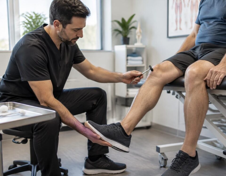
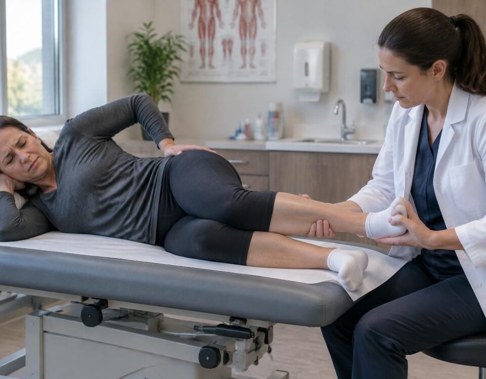
During the evaluation, the healthcare professional focuses on the onset of pain, movements that exacerbate it, numbness, and tingling. They also check strength, sensitivity, certain reflexes, and the ability to walk. Pain that radiates down the side of the leg or foot can guide the examination, but it does not, on its own, confirm which nerve root is irritated. The evaluation also considers other possible causes.
For a recent sciatica without alarming signs, an MRI is generally not the first step to take. The clinician may first gather history and conduct an examination. Imaging may be considered when pain persists, worsens, does not respond to initial measures, or when targeted treatment is discussed. The urgency of the examination depends on the clinical picture; the physician determines whether an MRI or another examination is appropriate.
This correlation between imaging and symptoms avoids treating a fortuitous finding as if it were necessarily responsible for the pain. It also helps to direct management towards the real needs of the individual, rather than solely following the radiological report. At TAGMED Clinic, the evaluation can guide a tailored non-surgical approach, including, depending on the situation, specific osteopathy, motorized neurovertebral decompression, or other modalities offered by the team. These treatments do not replace a diagnosis or medical follow-up.
Sylvain Desforges, B.Sc., D.O., N.D., doctor of osteopathy and naturopath, is the founding president of TAGMED and the ACMA association. His practice focuses especially on chronic spinal pain, lower back pain, and symptoms compatible with sciatica. He is interested in integrating technologies such as neurovertebral decompression, laser, and shockwaves, depending on the evaluation and care objectives. His approach aims to offer individualized support based on available data, without replacing authorized medical care.
A significant or progressive weakness, a new difficulty in walking, loss of control over the bladder or bowels, or numbness in the saddle area requires urgent medical evaluation. Severe and rapidly worsening pain, fever, or recent trauma also justify prompt consultation.
In Terrebonne, pain that radiates into the buttock or leg may suggest sciatica, but an MRI alone does not identify the cause. To guide management, the result must correspond to the pain’s path, changes in sensitivity or strength, and findings from the clinical examination.
Why MRI alone is not sufficient to explain pain
An MRI shows the structures of the spine: discs, nerve roots, and spaces through which the nerves pass. It may reveal a disc herniation or narrowing, but these signs do not automatically prove they are responsible for the symptoms. Some abnormalities also appear in individuals who are not experiencing pain. This is why imaging must be interpreted alongside the history of symptoms and the physical examination, as explained in this article about the relationship between disc herniation, MRI, and clinical examination.
Pain may also originate from nerve irritation without the image perfectly explaining its intensity or pathway. Conversely, a visible anomaly may be located at a level that does not correspond to the symptoms. To better understand this distinction, refer to the explanations about radicular pain compared to referred pain and the concordance between sciatica symptoms and MRI.
The pathway of symptoms helps to interpret the image
A lumbar condition can lead to pain that starts in the lower back and radiates into the buttock, thigh, leg, or foot. Tingling, loss of sensation, or weakness may accompany the pain. The affected area and movements that have become difficult provide clues to the healthcare professional, but they do not allow for a diagnosis on their own. A sensation of a heavy or painful leg, for example, deserves to be clarified in its context: heavy leg or painful leg in case of sciatica.
Some functional signs can guide the evaluation: difficulty standing on tiptoe may be examined in relation to a possible S1 root involvement, while loss of sensation in the toes must be described precisely. You can learn more about the S1 root and the difficulty in standing on tiptoe, as well as loss of sensation in the toes. These observations complement the examination; they do not replace a medical evaluation.
Compression at a foramen, the opening through which a nerve root exits the spine, may also be considered when symptoms and imaging coincide. To understand this notion, refer to this page on L5-S1 foraminal compression.
When to request an MRI and when to consult quickly
For recent sciatica without signs of severity, an MRI is not always immediately necessary. The professional first assesses symptoms, strength, sensitivity, reflexes, and mobility. Imaging may be considered if the pain persists, worsens, does not respond to initial measures, or if an injection or intervention is being considered. The indications depend on the individual situation; this guide presents guidelines on when an MRI may be useful for back pain, and this resource specifically addresses the sciatic nerve MRI, its indications, and what it can reveal.
An urgent consultation is necessary in cases of significant or progressive weakness, new difficulty walking, loss of urinary or bowel control, urinary retention, or numbness in the anal region. Extreme pain or rapid deterioration also justifies immediate medical advice. The recommendations on sciatica management emphasize the importance of identifying these signs before focusing on imaging results.
In Terrebonne, an evaluation provides perspective on the pain trajectory, its evolution, neurological signs, and, when available, the MRI. This approach helps avoid treating only a radiological anomaly that does not correspond to symptoms or neglecting a condition that requires medical advice. To know the evaluation options and the services offered, consult the page on sciatica treatment in Terrebonne.
About the author
Sylvain Desforges, B.Sc., D.O., N.D., Doctor of Osteopathy and Naturopath, is the founding president of TAGMED and the ACMA association. He dedicates his practice to chronic spinal pain, lower back pain, and pain associated with sciatica, particularly when the pain radiates down to the buttock, leg, calf, foot, or toes.
Through SOS Sciatique, he publishes educational content on sciatica symptoms, possible causes such as herniated discs, disc pinching, foraminal stenosis, or irritation of a lumbar nerve root, as well as non-surgical approaches that may be considered based on the evaluation. At TAGMED Clinic, care may include, depending on the case, motorized neurovertebral decompression, specific osteopathy, or precision percussion.
For an evaluation related to sciatica treatment in Montreal / Mont-Royal or sciatica treatment in Terrebonne, consult the available services at TAGMED Clinic.
Medical Disclaimer
The information published on SOS Sciatique is provided for educational purposes and does not replace a consultation, diagnosis, advice, or treatment by a doctor or another authorized health professional. The author of this article is neither a physician nor a specialist in a medical specialty recognized by the Collège des médecins du Québec. The osteopathy, manual medicine, or functional support approaches described on this site are not medical acts and are not intended to replace medical follow-up.
Consult a doctor or emergency service in case of loss of urinary or bowel control, anesthesia in the anal region, significant or progressive weakness of the leg or foot, fever, recent trauma, severe pain, or rapid deterioration. For more details, please read our Legal Notice.

Sylvain Desforges, B.Sc., D.O., N.D., docteur en ostéopathie
Dr Sylvain Desforges, B.Sc., D.O., N.D., osteopath, is the founding president of TAGMED and of the ACMA association. He dedicates his practice to chronic spinal pain, low back pain and symptoms compatible with sciatica, particularly when pain radiates toward the buttock, leg, calf, foot or toes.
Through SOS Sciatique, he publishes educational content on sciatica symptoms, possible causes such as disc herniation, disc narrowing, foraminal stenosis or irritation of a lumbar nerve root, as well as non-surgical approaches that may be considered according to the evaluation. At Clinique TAGMED, care may include, depending on the case, motorized neurovertebral decompression, specific osteopathic care or precision percussion.
For an evaluation related to sciatica treatment in Montreal / Mount Royal or sciatica treatment in Terrebonne, please consult the services available at Clinique TAGMED.
Medical disclaimer
The information published on SOS Sciatique is provided for educational purposes only and does not replace a consultation, diagnosis, opinion or treatment provided by a physician or another authorized healthcare professional. The author of this article is neither a medical doctor nor a specialist in a medical specialty recognized by the Collège des médecins du Québec. The osteopathic, manual medicine or functional support approaches described on this site do not constitute medical acts and are not intended to replace medical follow-up.
Consult a physician or emergency service in cases of loss of bladder or bowel control, anesthesia in the saddle area, significant or progressive weakness in the leg or foot, fever, recent trauma, severe pain or rapid worsening of symptoms. For more details, please read our Legal Notice.