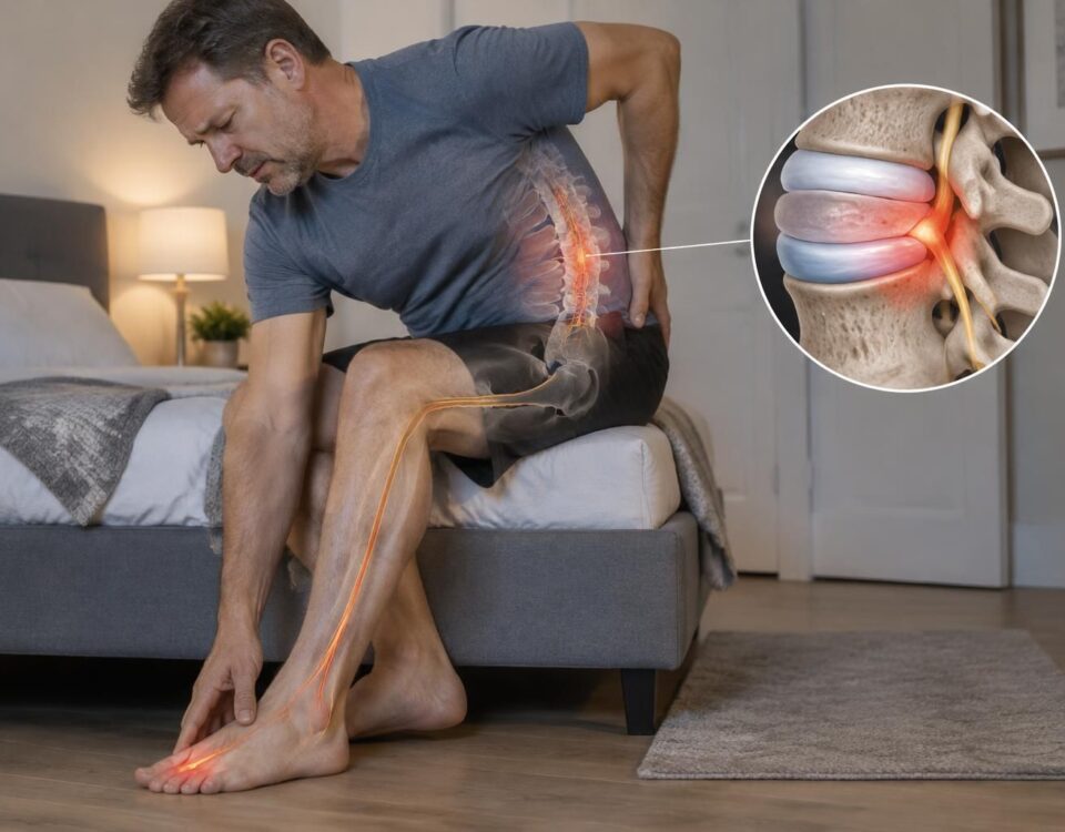



Sciatica and Chronic Pain: When to Reassess?
04/11/2026
Sciatica and pain during exercise: sign to watch out for
04/11/2026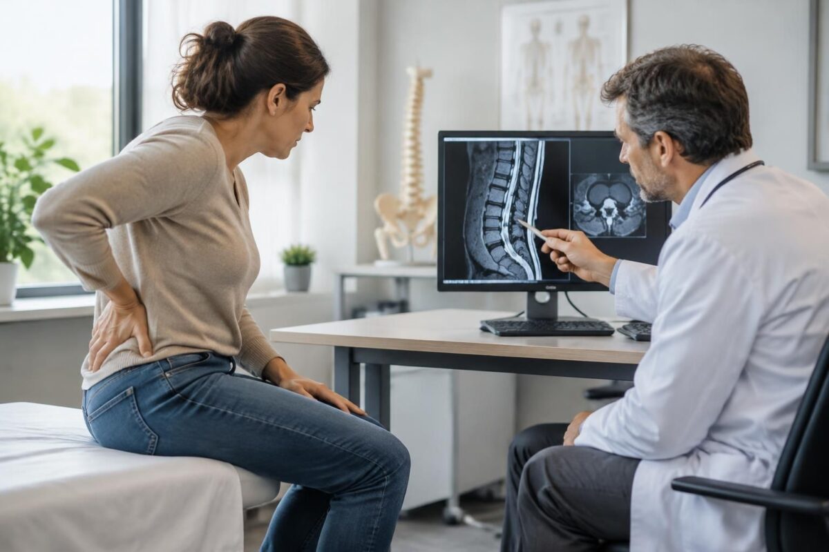
IN SHORT A disc bulge is a diffuse protrusion of a disc, sometimes associated with sciatica, but often discovered incidentally on imaging. To determine if it explains symptoms, one must compare the image to their path, the neurological examination, and functional limitations. This evaluation helps guide management without treating an isolated image. |
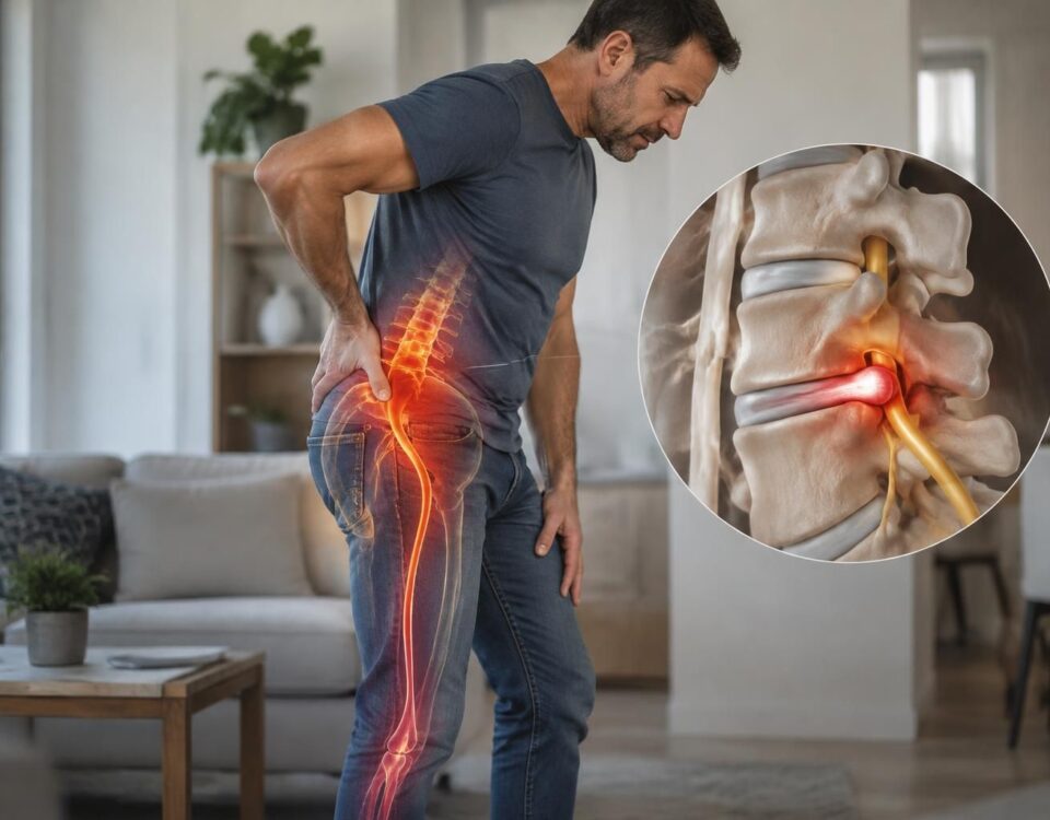
A disc bulge can contribute to sciatica if it corresponds to the side and path of the pain, the neurological examination, and functional limitations. However, this term describes a form observed on imaging: it does not prove, by itself, that the disc causes the symptoms.
Since some bulges are present without pain, their significance must be evaluated alongside the entire clinical picture. A progressive weakness, foot drop, loss of sensation in the saddle area, or urinary or bowel issues require urgent assessment.
A disc bulge can contribute to sciatica, but its presence on an MRI does not prove it explains the pain. To understand its implications, one must compare the image to the symptoms, their path, the neurological examination, and their progression. This distinction helps avoid treating a term from the report rather than the actual situation. The disc is located between two vertebrae. With age, repeated stresses, and individual characteristics, its contour may change. A disc bulge generally refers to a fairly extensive protrusion of the disc’s contour. A disc protrusion rather describes a localized bulge whose base is broader than its depth. These terms describe different forms and are not interchangeable. Imaging reports may also mention extrusion or sequestration. These words indicate the shape and continuity of the disc material. They do not, by themselves, determine the intensity of pain, the affected nerve root, or the appropriate treatment. To delve deeper into possible mechanisms, refer to this guide on the causes and degeneration of bulges or protrusions, as well as this resource on disc bulges and their possible causes. Yes, in some cases. A disc protrusion can reduce the space around a nerve root or irritate surrounding tissues. When the symptoms and examination correspond to that root, the bulge may be a plausible cause of sciatica. However, an abnormal image may also be a finding with no direct link to the symptoms. People without pain sometimes present disc changes on imaging. Sciatica refers to pain that follows part of the path of the sciatic nerve or a lumbar nerve root. It can start from the lower back or buttock and travel down the thigh, leg, or foot. Some people also experience numbness, tingling, or weakness. A bulge or protrusion associated with radiculopathy can only be considered as an explanation after an assessment that verifies the concordance between symptoms and clinical signs. The healthcare professional inquires about the onset of the symptoms, their duration, their side, and their path. They also ask which positions or activities worsen or relieve them. Pain in the hip, for instance, does not necessarily mean that a nerve is irritated. This guide helps distinguish hip pain from true nerve irradiation. The area of pain can guide the evaluation, but it is not sufficient to make a diagnosis. Irritation of the L5 root may sometimes be accompanied by symptoms on the top of the foot or towards the big toe. Irritation of S1 may affect the back of the leg and the outer edge of the foot. These paths vary from person to person. For more information, consult the pages on pain on the top of the foot, pain in the big toe, and possible signs of an L4-L5 protrusion. The examination can compare strength, sensation, reflexes, and walking. The professional also checks movements and activities that matter to the person. A weakness, loss of sensation, or change in function may be more significant than an isolated change in pain. The decision takes into account the entire picture: symptoms, examination, function, and imaging when relevant. A resource on lumbar bulges, stenosis, and symptoms in the leg on MRI can help understand certain terms but does not replace clinical interpretation. For a general overview of the topic, you may also read this page on disc bulging. An X-ray and an MRI do not answer the same questions. The X-ray mainly shows the bony structures and alignment. The MRI allows visualization of the discs and the tissues around the nerves. The choice of an exam depends on the symptoms, examination results, and the clinical question posed. Imaging is not always necessary, especially when symptoms are stable and no concerning signs are present. It may be considered if symptoms persist, if a deficit arises, if another diagnosis is possible, or if the result may change management. The professional determines if an examination is indicated; an MRI report should not be interpreted in isolation. Pain radiating down the leg can have several explanations. It may result from nerve irritation, but also from a joint, muscle, or another structure. Hip pain may sometimes resemble sciatica. Some less common causes, such as infection, fracture, or systemic illness, require appropriate medical evaluation. Information about the onset of pain, recent trauma, fever, general health status, and history helps guide the evaluation. Persistent pain in the leg merits re-evaluation if it changes, worsens, or is accompanied by new signs. You can consult this guide on disc protrusion and persistent pain in the leg. An effective plan starts with a concrete goal: improving walking, resuming an important activity, sleeping better, or reducing a limitation at work. The professional notes the symptoms and relevant neurological elements and sets a date for re-evaluation. They also specify which changes would warrant modifying the plan or seeking another opinion. The options depend on the diagnosis and the situation. A non-surgical approach may be considered for certain profiles, but no method is suitable for everyone and no results can be guaranteed. Neurovertebral decompression, specific osteopathy, or other treatments should be discussed after an assessment. They do not replace a medical consultation when a diagnosis or medical follow-up is necessary. The TAGMED Clinic offers services in Montreal and Terrebonne. Readers can consult information about sciatica treatment in Montreal and sciatica treatment in Terrebonne. The TAGMED Clinic and The Spine Page belong to the same portfolio; these clinical pages describe a service offering and do not replace a comparison of options or urgent care when required. A new or progressive weakness, a dropped foot, increasing difficulty walking, loss of dexterity, or numbness that spreads requires rapid evaluation. Loss of control of the bladder or bowels, or loss of sensation in the saddle area, demands urgent evaluation. Severe or rapidly worsening pain, fever, or pain following a recent trauma also justifies a quick medical opinion. If these signs are present, do not delay evaluation to complete a treatment protocol. No. It is necessary to check if the level and side of the anomaly correspond to the symptom trajectory, neurological examination, and evolution. Imaging alone does not confirm the cause of the pain. No. A bulge generally describes a more diffuse extension of the disc, while protrusion corresponds to a more localized protrusion. These terms characterize the observed shape and do not alone determine the presence of symptoms. Not reliably. A large anomaly may not cause pain, while a more discreet change may be relevant if it aligns with the symptoms and examination. No. An MRI is particularly useful if its result can change the management, if symptoms persist, or if a deficit or other concerning sign appears. The professional assesses the necessity of the examination. Not necessarily. Pain, strength, sensitivity, and walking can evolve differently. Weakness or loss of function deserves particular attention, even if the pain decreases. Sylvain Desforges, B.Sc., D.O., N.D., Doctor of Osteopathy and Naturopath, is the founding president of TAGMED and the ACMA association. His practice focuses on chronic pain of the spine, lower back pain, and pain compatible with sciatica, particularly when the pain radiates down to the buttock, leg, calf, foot, or toes. Through SOS Sciatica, he publishes educational content about sciatica symptoms, possible causes—including herniated disc, disc pinching, foraminal stenosis, or irritation of a lumbar nerve root—and non-surgical approaches that may be considered based on the evaluation. At the TAGMED Clinic, care may include, depending on the case, motorized neurovertebral decompression, specific osteopathy, or precision impactor. Information published on SOS Sciatica is provided for educational purposes and does not replace a consultation, diagnosis, opinion, or treatment by a physician or other licensed healthcare professional. The author of this article is neither a doctor nor a specialist in a medical specialty recognized by the Collège des médecins du Québec. The osteopathy, manual medicine, or functional support approaches described on this site do not constitute medical acts and are not intended to replace medical follow-up. Consult a physician or emergency service in case of loss of bladder or bowel control, anesthesia in the saddle region, significant or progressive weakness of the leg or foot, fever, recent trauma, severe pain, or rapid worsening. For more details, please read our Legal Notice. Medical disclaimer: The information and advice provided on this site do not replace the opinion, diagnosis, or treatment of a healthcare professional. Please note that Sylvain Desforges, B.Sc., D.O., N.D., Doctor of osteopathy, is neither a medical doctor nor a physician, and is not a specialist in a medical specialty as defined by the Collège des médecins du Québec. Manual medicine, functional medicine, and sports medicine as described on this site exclude any treatment or medical diagnosis made by a physician or specialist doctor. Always consult your physician for any medical questions. For more details, please read our complete Legal Notice.What does “disc bulge” mean on an MRI?
Can a disc bulge cause sciatica?
How to check if the image corresponds to the symptoms?
The path of the pain
The neurological examination and function
When is imaging useful?
What other causes should be considered?
How to establish a management plan?
What signs require rapid or urgent evaluation?
Frequently asked questions about disc bulge and sciatica
Does a disc bulge always explain sciatica?
Is a protrusion the same as a bulge?
Does the size of the anomaly indicate the intensity of the pain?
Is an MRI always necessary?
Does decreasing pain mean the nerve has recovered?
About the author
Medical Warning
When the bulge may be responsible
- Pain radiating into the leg, compatible with a nerve root.
- Side and level of the MRI correlating with the symptoms.
- Examination showing changes in strength, sensitivity, or reflexes.
- Evolution of symptoms consistent with the clinical picture.
When it may be incidental
- Finding on imaging without corresponding symptoms.
- Level or side that does not match the pain.
- Symptoms better explained by another cause.
- The term “bulge” alone does not determine treatment.
To verify: symptoms, function, neurological examination, and imaging should be considered together.
Urgency: progressive weakness, dropped foot, saddle anesthesia, or urinary or bowel disturbances require rapid evaluation.

A disc bulge can contribute to sciatica when it irritates a nerve root and its location correlates with the symptoms. But this finding on imaging can also be an incidental discovery unrelated to the pain. To interpret it, the MRI report must be correlated with the symptoms, neurological examination, and their evolution, rather than treating an isolated term.
Disc bulge and protrusion: different terms
The bulge generally describes a relatively wide extension of the contour of the disc. The disc protrusion refers more to a localized herniation, where the base is wider than the part that protrudes. These terms describe the observed shape: they do not alone determine if a person is in pain, which nerve root is involved, or what treatment would be appropriate.
Bulges and protrusions can be present in people who have no symptoms. Conversely, pain in the buttock or leg can have other causes. The size of the anomaly does not always predict the intensity of the pain. The essential question is therefore that of the concordance between the image and the clinical picture.
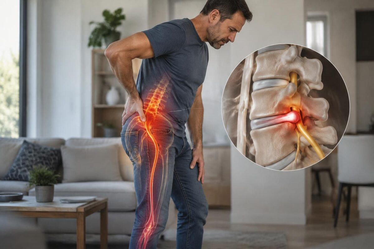
Disc herniation L4-L5 and leg pain
IN BRIEF A L4-L5 disc bulge is a protrusion of the lumbar disc that can contribute to pain in the buttock or leg if a nerve root is irritated. An MRI in a neutral position does not always show what…

Lumbar Disc Protrusion and the Choice of a Non-Surgical Approach
IN SHORT A disc protrusion is a bulging of a disc between the vertebrae; it often does not cause any symptoms. If it is accompanied by pain, a non-surgical approach may include appropriate activity, prescribed medications, and progressive rehabilitation. It…
When can the bulge be related to sciatica?
Compare the symptom trajectory
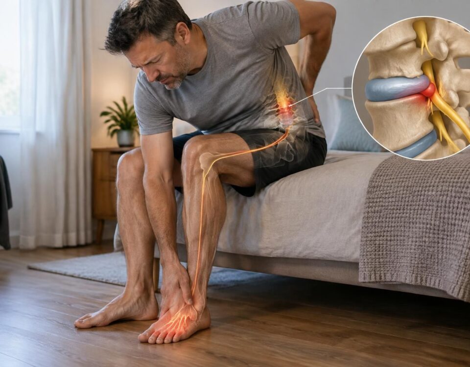
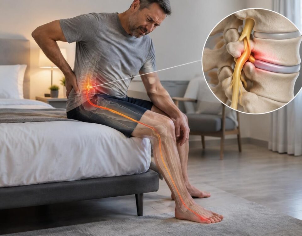
Sciatica can manifest as pain starting from the lower back or buttock and descending into the leg, sometimes down to the foot or toes. Tingling, numbness, or weakness may also occur. The location and side of the symptoms should be compared to the level and side described in the imaging report. This comparison helps the professional determine if the disc could be contributing to the symptoms, but is not sufficient to establish a diagnosis.
Consider the examination and function
The evaluation may include the history of symptoms, activities that worsen or relieve them, walking, as well as strength, sensitivity, and reflexes. It is also useful to note the impact on sleep, work, and daily activities. An image that does not correspond to either the painful territory or the observations of the examination could be an incidental finding.
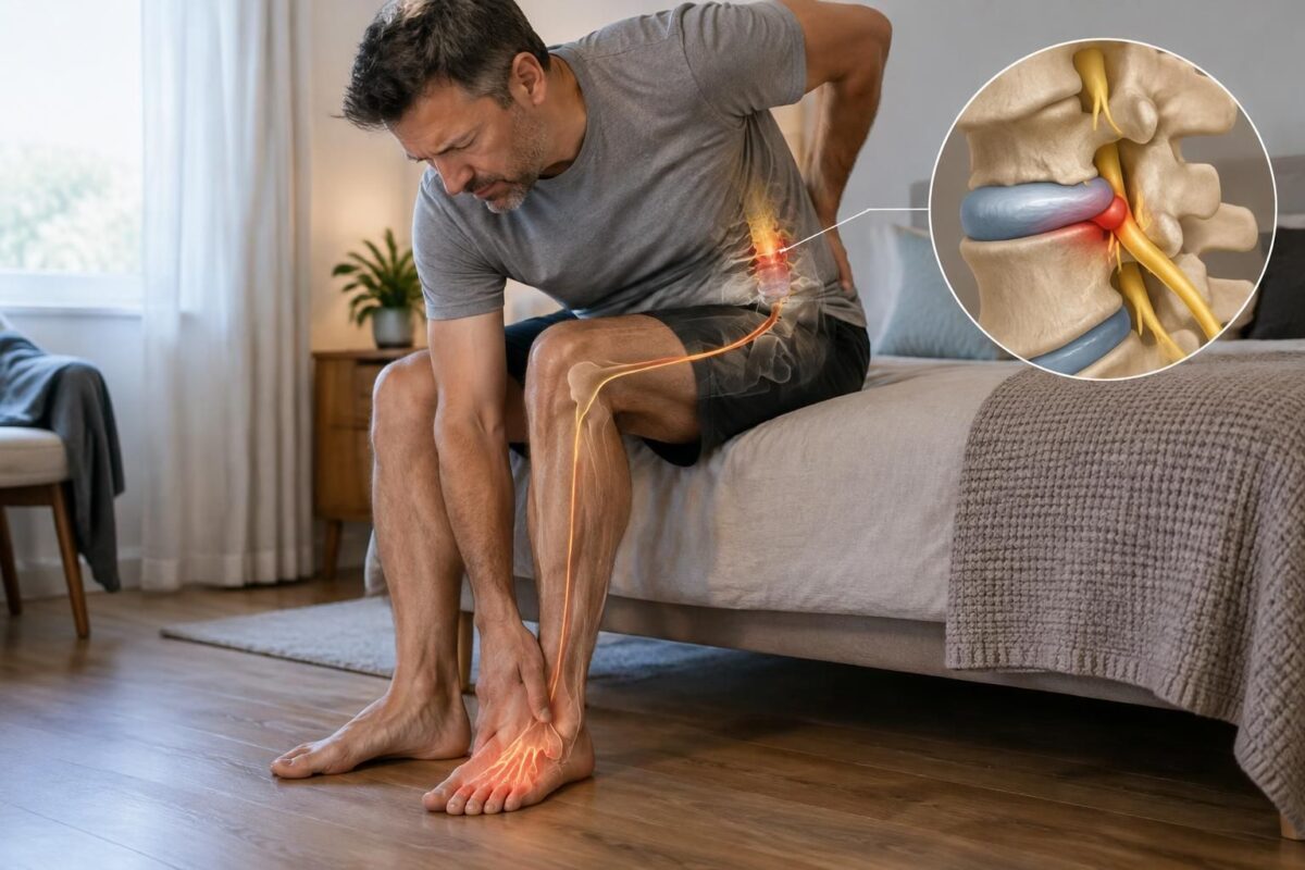
Disc herniation and pain on the top of the foot
IN BRIEF A disc protrusion is an intervertebral disc that bulges without necessarily rupturing. Pain on the top of the foot may sometimes be related to irritation of a lumbar nerve, but it does not alone confirm disc involvement. An…
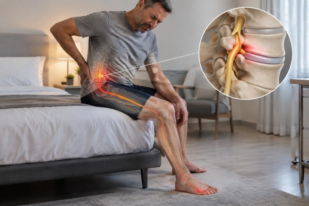
Disc protrusion and pain on the outer edge of the foot
IN BRIEF A disc bulge is a bulging of the disc that can sometimes irritate a nerve root and cause pain down to the outer edge of the foot. However, this pain is not always related to the disc: a…
What to do after an MRI result?
Avoid drawing a conclusion from a single term in the report. Ask the professional accompanying you what the image may explain, what other causes are possible, and whether additional examinations could change the management. Imaging is not automatically necessary or useful in every situation: its relevance depends on the symptoms, the examination, and the clinical decision to clarify.
If the symptoms are stable, a graduated plan may be proposed based on your situation. Set a concrete goal with the professional — for instance, walking longer or sleeping better — as well as a date for reevaluation. Follow-up should consider the evolution of pain, but also function and neurological signs. A temporary improvement alone does not establish the cause of the problem.
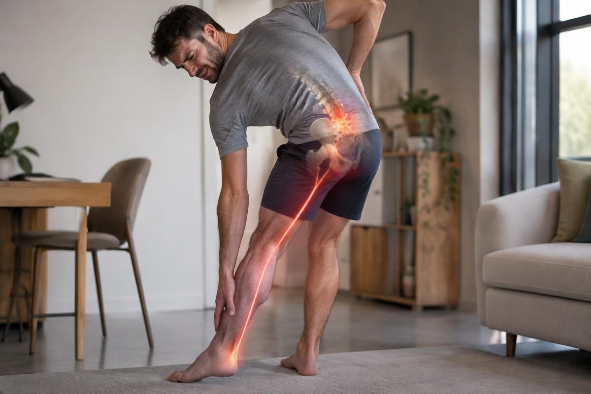
IN BRIEF A disc herniation is a bulging of a disc in the spine, often without symptoms. If it irritates a lumbar nerve root, the pain may radiate down the leg to the foot and sometimes be felt in the…

Intervertebral disc protrusion and pain in the big toe
IN BRIEF A disc protrusion is a bulging of the disc that can irritate a lumbar nerve root, particularly L5, and cause pain, tingling, or numbness down to the big toe. This symptom alone does not confirm a disc origin:…
When to request a rapid evaluation?
Consult a professional promptly if weakness appears or progresses, if the foot begins to drop, if numbness spreads, or if you have new difficulties walking. Loss of bladder or bowel control, or numbness in the saddle region, requires urgent evaluation. In the presence of significant neurological change, do not delay evaluation to continue a care program.
This information is educational and does not replace a consultation, diagnosis, or opinion from a licensed health professional.
| Compared element | Indicator to check |
|---|---|
| Location | The side and level concord with the pain trajectory. |
| Symptoms | The pain descends into the buttock or leg; a bulge alone is not sufficient to explain sciatica. |
| Neurological examination | Strength, sensitivity, and reflexes may reveal a compatible impairment. |
| Function | Walking, sleep, or activities are consistently limited in relation to the symptoms. |
| Evolution | The onset and changes of symptoms help assess the relevance of the imaging result. |
| Incidental finding | Possible if the image does not conform to the symptoms and examination. |
| Recommended action | The clinical evaluation guides the follow-up; the image alone does not determine treatment. |
| Signs of urgency | Progressive weakness, saddle anesthesia, or urinary or bowel disorders: urgent evaluation. |
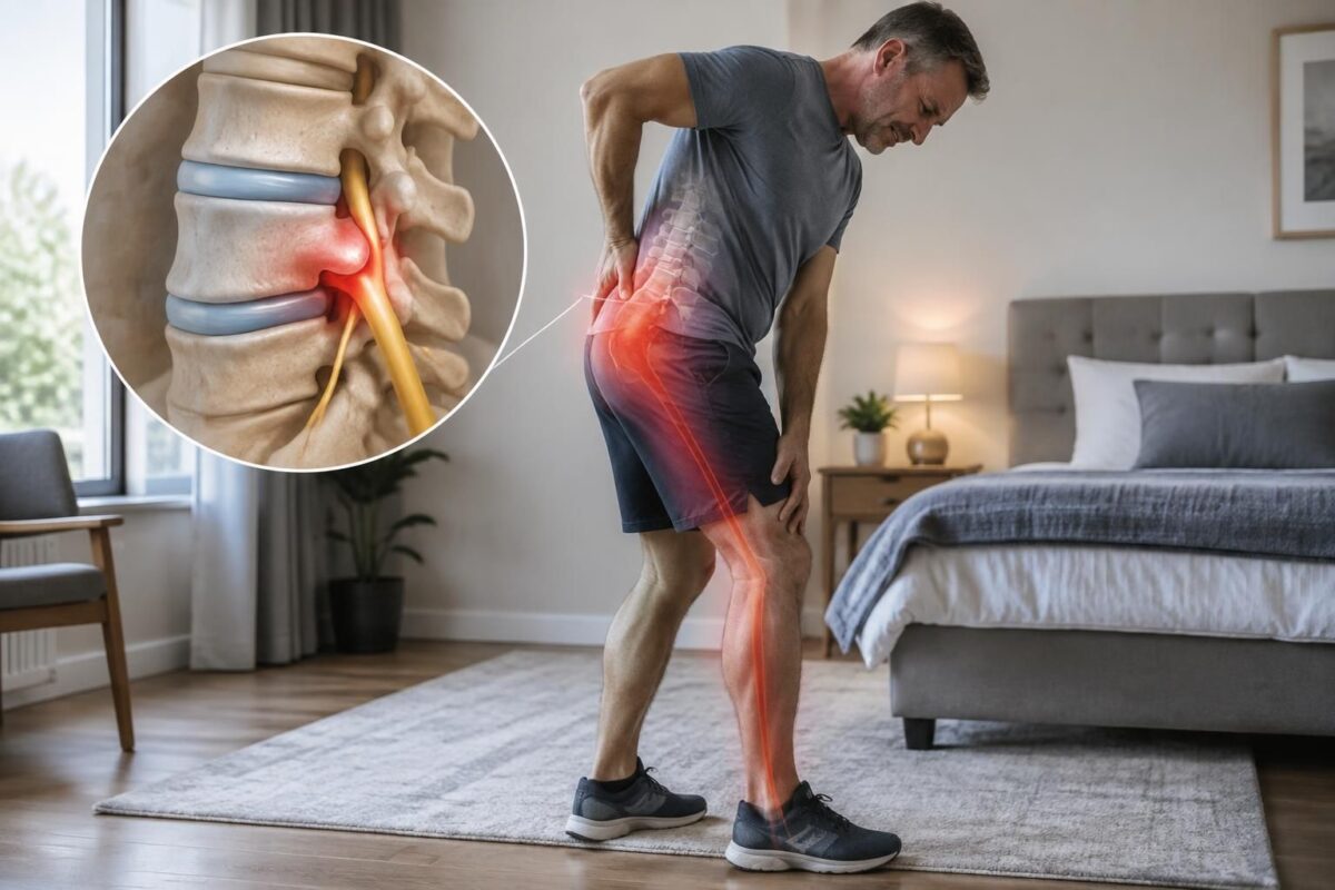
Disc herniation and pain radiating below the knee
IN BRIEF A disc protrusion is a deformation of a disc in the spine that can irritate a nerve root. If the pain extends below the knee, with tingling or weakness, this may suggest sciatica, although this alone does not…
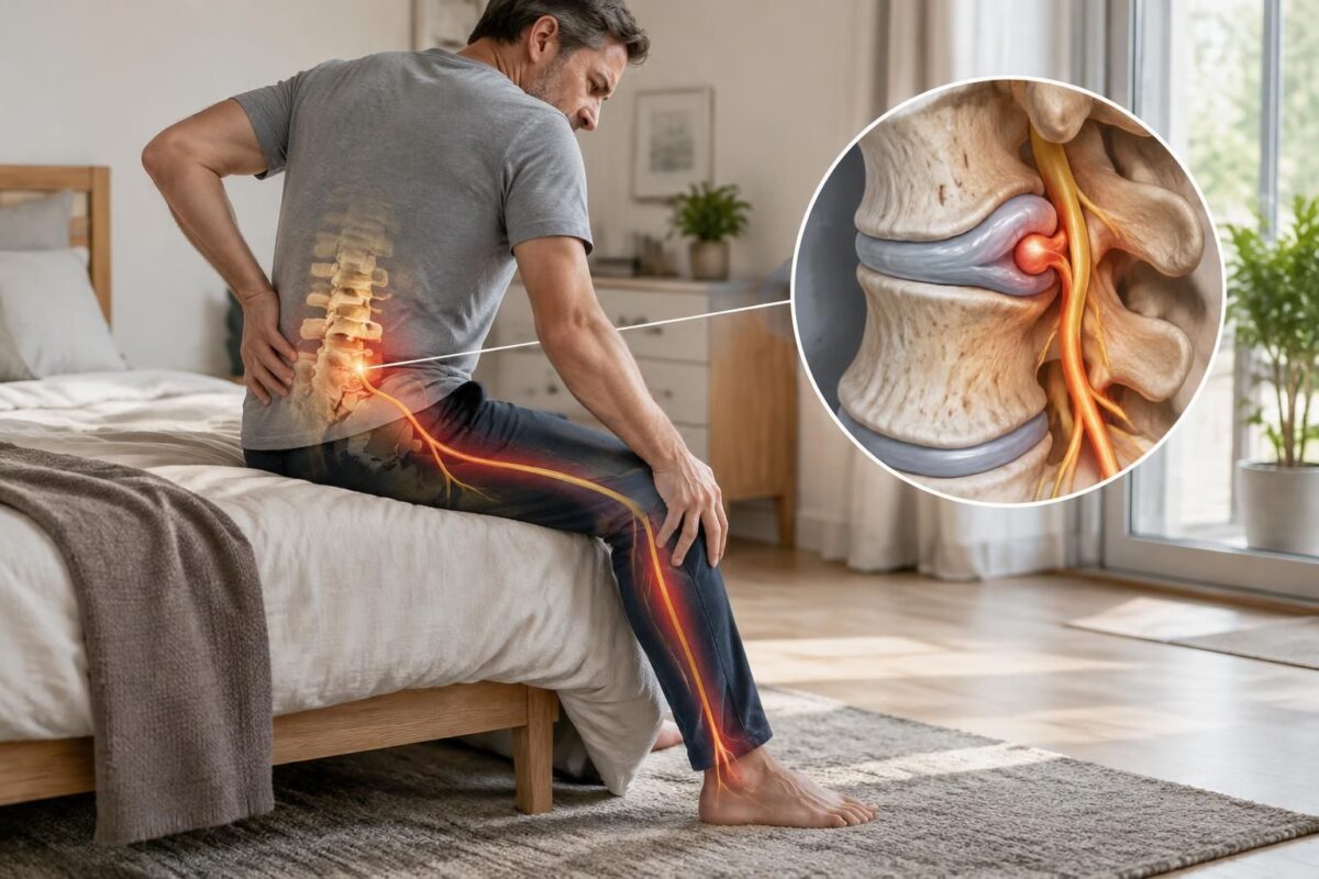
Intervertebral disc protrusion and neurological symptoms
IN BRIEF A disc protrusion is an intervertebral disc that bulges and can irritate a nerve root. It can cause pain, tingling, numbness, or weakness in an arm or leg, depending on its location. Clinical examination and, if necessary, imaging…
Disc bulge and sciatica: illustrative testimonies
Transparency note: The stories below are fictional and serve to illustrate various possible situations. They do not correspond to actual patient testimonies and do not allow for a diagnosis.
“The MRI showed a bulge, but my symptoms did not match”
“I had lower back pain and was worried when reading the report of my MRI. It mentioned a disc bulge, so I thought it must be the cause of my pain. During the evaluation, I was explained that it was also important to consider where the pain was radiating, the activities that modified it, and the results of the clinical examination. I understood that an image alone is not sufficient to establish the origin of the symptoms.”
“The pain in my leg matched the evaluation”
“My pain started in the lower back and descended into the buttock, then towards the leg. The report described a disc anomaly on the same side. The professional compared this result to my pain territory, my strength, and my sensitivity. I was informed that this concordance could make the result relevant, without the term “bulge” automatically confirming the cause.”
“I learned that bulge and protrusion do not refer to the same shape”
“I thought that “bulge” and “protrusion” were two equivalent terms. I was explained that they describe different morphologies of the disc. I also learned that these imaging words alone do not indicate whether a person will experience pain or what treatment will suit them.”
“My progress was not just about the pain”
“The pain varied from day to day. To track the evolution, I also noted how long I could walk, how I was sleeping, and if there were changes in strength or sensitivity. This approach helped me discuss concrete results rather than draw conclusions from just one day or one number.”
“A new symptom changed the priority”
“Initially, I focused on the imaging report. Then, a progressive weakness in my foot appeared. I was informed that this change needed to be evaluated quickly, regardless of the size of the bulge described. I understood that a neurological deterioration should not wait for the next step of a care program.”
These examples remind us that a useful assessment correlates symptoms, function, neurological examination, and imaging when relevant. A progressive weakness, a dropped foot, anesthesia in the saddle area, or urinary or bowel disturbances require urgent medical evaluation.
When imaging reveals a disc bulge in someone suffering from sciatica, it is natural to wonder if the two are related. The answer depends on the context: this disc change may contribute to the irritation of a nerve root, but it may also be an incidental finding. Therefore, the imaging report alone is not sufficient to identify the source of the pain or to determine the appropriate treatment.
A bulge generally corresponds to a fairly diffuse extension of the contour of the intervertebral disc. It differs from a disc protrusion, which refers to a more localized herniation, whose base is wider than its depth. The terms “protrusion,” “extrusion,” and “sequestration” describe different forms and continuities of disc material. They are not interchangeable, but their appearance alone does not predict the intensity of symptoms.
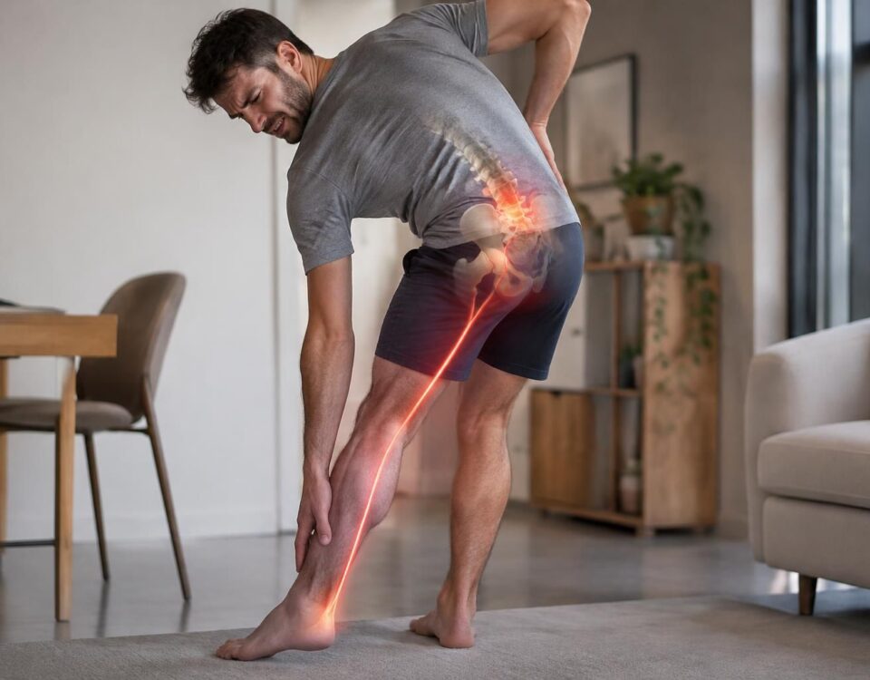
Sciatica often manifests as pain that starts from the lower back or buttock and extends down the leg. It may be accompanied by tingling, numbness, or sometimes weakness. The distribution varies according to the affected nerve root. For example, pain related to L5 may reach the side of the leg and the top of the foot, while an S1 involvement may extend to the back of the leg and the outer edge of the foot. These pathways provide clues but do not alone establish a diagnosis.
To assess whether the observed bulge is relevant, clinical correlation must be checked: do the level and side described in imaging correspond to the pathway of the symptoms, the limited activities, and the exam findings? The evaluation may consider strength, sensitivity, reflexes, walking, and movements that worsen or relieve the pain. A visible bulge in an asymptomatic person, or located at a level that does not match the signs, may not explain the sciatica.
Age, disc degeneration, repetitive strain, and anatomy can contribute to the onset of disc changes. However, imaging does not automatically designate an event or activity as the cause. An X-ray and an MRI address different questions; imaging is not necessary in all cases. It is primarily considered when its results can change management, in the presence of concerning signs, or before a targeted procedure.
Follow-up benefits from measuring factors beyond pain alone: ability to walk, sleep, effort tolerance, work participation, and changes in neurological signs. A care plan should specify a functional goal, a trial period, and a re-evaluation date. Depending on the situation, non-surgical approaches may be discussed with a qualified professional. Neurovertebral decompression, like any intervention, is not suitable for everyone and does not replace a proper assessment.
A progressive weakness, a dropped foot, numbness in the saddle region, or urinary or bowel disturbances require swift or urgent medical evaluation. The same applies in cases of significant worsening or new neurological signs.
About the author: Sylvain Desforges, B.Sc., D.O., N.D., is an osteopath and naturopath. Founder and president of TAGMED clinics and the ACMA association, he dedicates his practice to managing chronic pain, particularly spinal pain. His approach may integrate, depending on the evaluation and context, technologies such as neurovertebral decompression, laser, or shock waves.
Disclaimer: this content is provided for educational purposes and does not replace consultation, diagnosis, or treatment by a licensed healthcare professional.
A disc bulge may contribute to sciatica, but it may also be an incidental finding on imaging. To determine whether it is relevant, one must compare its location and side to the symptoms, the neurological examination, and functional limitations; the term recorded in a report alone does not establish the cause of the pain or the appropriate treatment.
Assessing the Relevance of the Disc Bulge
A bulge generally corresponds to a fairly diffuse extension of the disc contour. A disc protrusion, on the other hand, refers to a localized herniation whose base is wider than its depth. These morphological descriptions are not interchangeable, and neither automatically proves that a nerve is irritated. To clearly distinguish these terms, refer to the guide on the difference between disc herniation and disc bulge as well as this presentation of disc bulge.
Clinical correlation is essential. Low back pain that radiates into the buttock and leg may suggest nerve irritation, but its pathway, evolution, and associated signs must be evaluated. Pain toward the back of the foot or the big toe may direct the exam differently than pain that follows the back of the leg down to the foot. Examples of symptoms associated with L4-L5 involvement are presented in this case of L4-L5 protrusion and leg pain; calf pain that appears while walking also requires careful evaluation, as explained in this guide on sciatica and calf pain while walking.
An MRI shows anatomical structures; it does not alone determine whether an anomaly is responsible for the symptoms. Some bulges or protrusions are present in individuals who feel no pain. Therefore, interpretation benefits from considering the affected side, painful territory, strength, sensitivity, reflexes, and activities that have become difficult. To better understand what the exam can reveal, see what the MRI can show in cases of disc herniation and sciatica, as well as information on the L4-L5 lumbar disc.
A useful assessment specifies the onset and duration of symptoms, their distribution, the movements or efforts that alter them, and the care already attempted. It also examines gait, mobility, strength, and sensitivity, then establishes baseline measures: for example, the tolerated walking distance, sleep, or an important daily task. Imaging is not necessary in all situations; it is more considered when it can change management, in the presence of concerning signs, or before a targeted procedure. General resources on the link between herniated disc and sciatica and sciatica can supplement this information without replacing an individual assessment.
Management depends on the clinical picture, evolution, and the person’s goals. A plan should specify what will be monitored, the duration of the trial, and when it will be reviewed. A non-surgical approach may be discussed in some cases, without the promise of universal results; its objectives, limitations, and risks must be explained. To compare options, consult the guide on the choice of a non-surgical approach and the information from this resource dedicated to symptoms and treatments of disc bulge. These external resources are informative and do not replace professional advice.
An improvement in pain is not always sufficient to confirm a complete recovery: strength, sensitivity, gait, sleep, and participation in activities may evolve differently. Conversely, progressive weakness, a dropped foot, loss of dexterity, anesthesia in the saddle region, or urinary or bowel issues require a rapid or urgent medical assessment. In the presence of such signs, do not delay referral to pursue a care program.

Sylvain Desforges, B.Sc., D.O., N.D., docteur en ostéopathie
Dr Sylvain Desforges, B.Sc., D.O., N.D., osteopath, is the founding president of TAGMED and of the ACMA association. He dedicates his practice to chronic spinal pain, low back pain and symptoms compatible with sciatica, particularly when pain radiates toward the buttock, leg, calf, foot or toes.
Through SOS Sciatique, he publishes educational content on sciatica symptoms, possible causes such as disc herniation, disc narrowing, foraminal stenosis or irritation of a lumbar nerve root, as well as non-surgical approaches that may be considered according to the evaluation. At Clinique TAGMED, care may include, depending on the case, motorized neurovertebral decompression, specific osteopathic care or precision percussion.
For an evaluation related to sciatica treatment in Montreal / Mount Royal or sciatica treatment in Terrebonne, please consult the services available at Clinique TAGMED.
Medical disclaimer
The information published on SOS Sciatique is provided for educational purposes only and does not replace a consultation, diagnosis, opinion or treatment provided by a physician or another authorized healthcare professional. The author of this article is neither a medical doctor nor a specialist in a medical specialty recognized by the Collège des médecins du Québec. The osteopathic, manual medicine or functional support approaches described on this site do not constitute medical acts and are not intended to replace medical follow-up.
Consult a physician or emergency service in cases of loss of bladder or bowel control, anesthesia in the saddle area, significant or progressive weakness in the leg or foot, fever, recent trauma, severe pain or rapid worsening of symptoms. For more details, please read our Legal Notice.