


Severe sciatica: what signs warrant an evaluation?
09/11/2026
Sciatica and prolonged immobility: possible effect on pain
09/11/2026
IN BRIEF
| A disc bulge is a protrusion of the disc between the vertebrae, which may not necessarily cause symptoms. When pain fluctuates, its intensity can vary based on movements, activities, and other factors; the bulge visible on MRI does not always explain what one feels on its own. Evaluating symptoms and conducting a clinical examination helps determine the next steps and appropriate non-surgical options. Consult quickly if there is worsening weakness or loss of control of the bladder or bowels. |
A disc bulge can be associated with pain that varies with movements, postures, or levels of activity. Tingling, numbness, or pain radiating into an arm or leg may also fluctuate, especially when a nerve is irritated. However, a bulge seen on an MRI does not necessarily explain the symptoms: its significance must be assessed based on the clinical examination and their location.
If symptoms persist or worsen, seek the advice of a healthcare professional. Progressive weakness, loss of bladder or bowel control, or numbness in the saddle area requires urgent evaluation.
A disc bulge may be associated with pain that varies over days, activities, or positions. This fluctuation does not necessarily mean that the disc is changing rapidly. The MRI should be interpreted alongside the symptoms and clinical examination, as a mild bulge can also be present without causing pain.
What does a disc bulge mean?
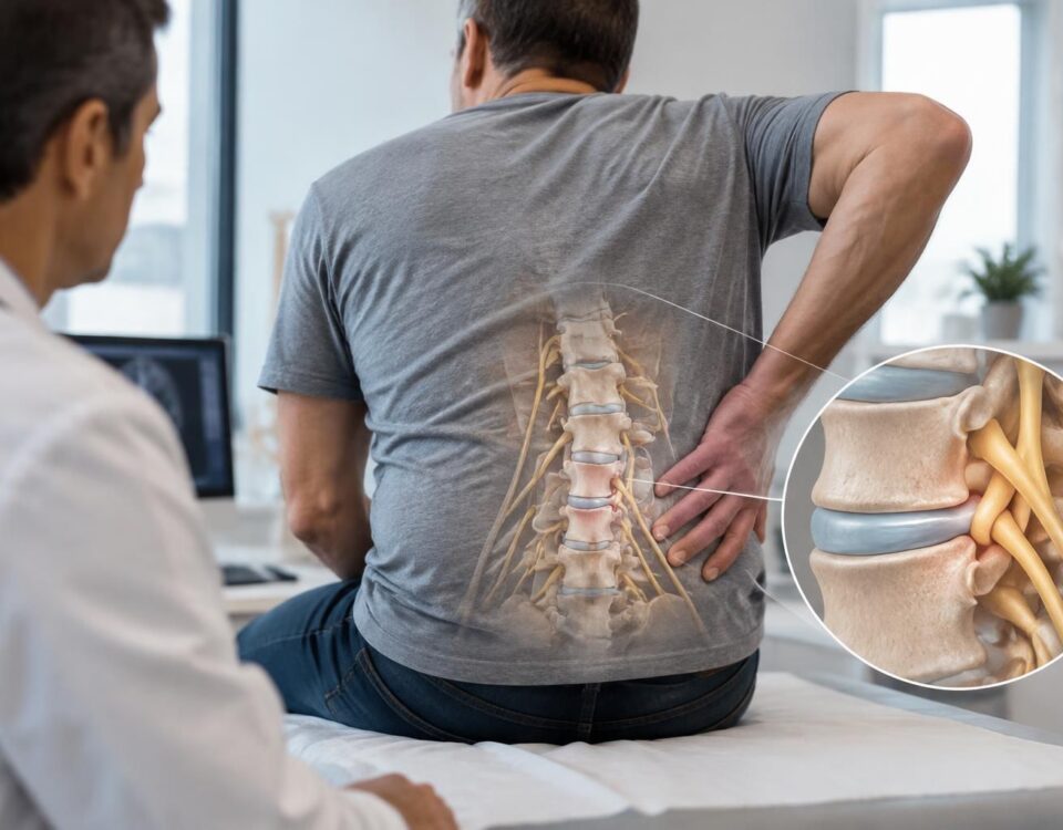
Intervertebral discs are located between the vertebrae. They help absorb stress and allow the spine to move. A bulge describes a border of the disc that exceeds its usual boundary, often over a fairly large area. This term does not necessarily mean that there is a rupture of the disc or compression of a nerve.
A disc bulge is different from a herniated disc. A herniation corresponds to a more localized protrusion of disc material. However, the terms used in an imaging report are not sufficient to determine the cause of pain. To better understand what the MRI can show, consult this article on herniated discs and sciatica on MRI.
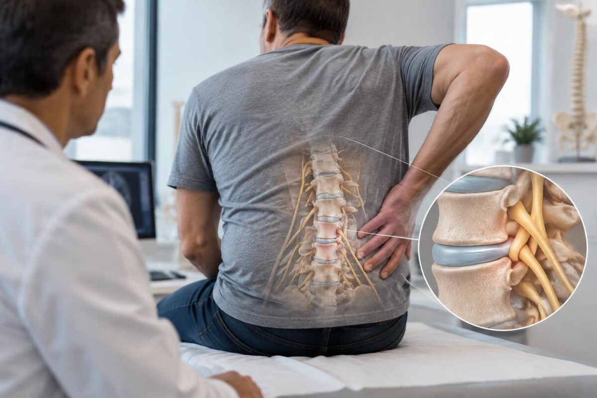
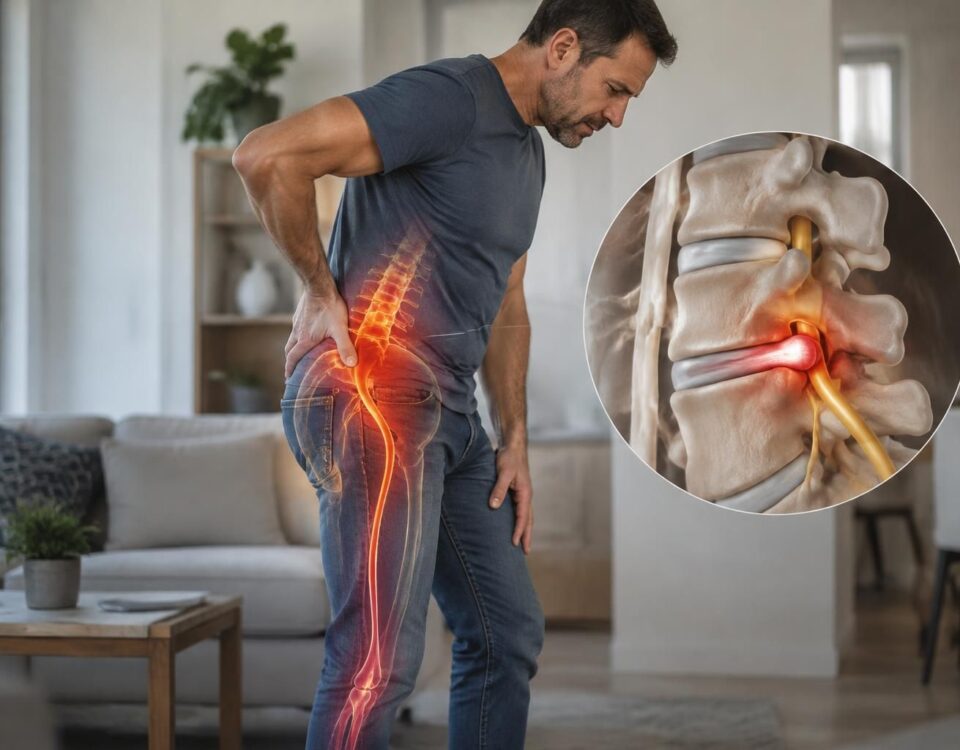
Disc bulge and mild nerve compression
IN BRIEF A disc bulge with mild nerve compression occurs when a disc slightly protrudes and may irritate a nerve, sometimes causing pain, tingling, or numbness. Symptoms and imaging do not always align: the assessment takes into account your movements…
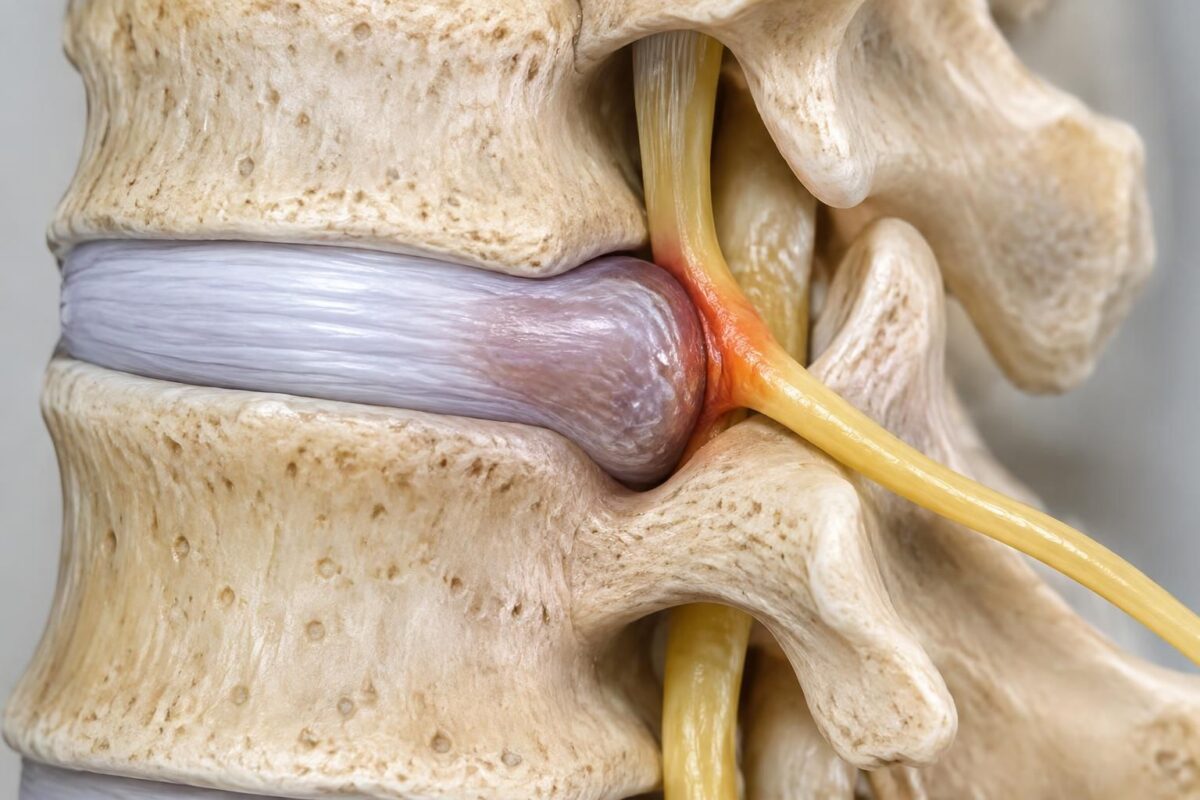
Disc herniation and foraminal irritation
IN BRIEF A disc bulge can reduce the space of the foramen, the opening through which a nerve exits, and contribute to its irritation. This can cause lower back pain radiating into the buttock or leg, tingling, or numbness. An…
Why can symptoms fluctuate?
Pain can increase or decrease without any major visible change in the disc. Daily activities, prolonged periods in the same position, sleep, a recent injury, and overall condition can influence symptoms. The sensitivity of muscles, joints, or nerves can also vary.
A movement or posture can sometimes trigger a temporary increase in pain. For example, some people notice discomfort after twisting their back. This reaction does not, by itself, conclude that a bulge has worsened. The article on pain after twisting the back presents factors to consider.
A pain that appears and then subsides
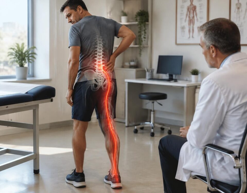
Intermittent pain may remain localized in the back or neck. It may also radiate to a buttock, leg, shoulder, or arm, depending on the affected area. Tingling or numbness may accompany certain radiating pains. These signs should be evaluated in context, especially if they persist or worsen.
Recurring symptoms
Repeated episodes do not prove that the disc bulge is enlarging. Symptoms may return when a sensitive area is further engaged or when the individual recovers less well. A recurrent sciatica warrants evaluation if it limits activities or if neurological signs progress.
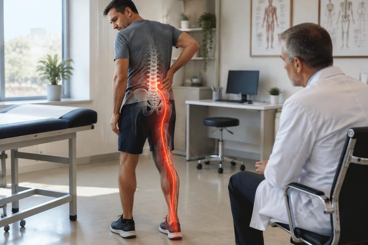
Disc bulge L5-S1 and pain toward the foot
IN SHORT A L5-S1 disc bulge corresponds to a disc that slightly overruns between the last lumbar vertebra and the sacrum. It can irritate a nerve root and cause pain resembling sciatica down to the foot, but this anomaly visible…
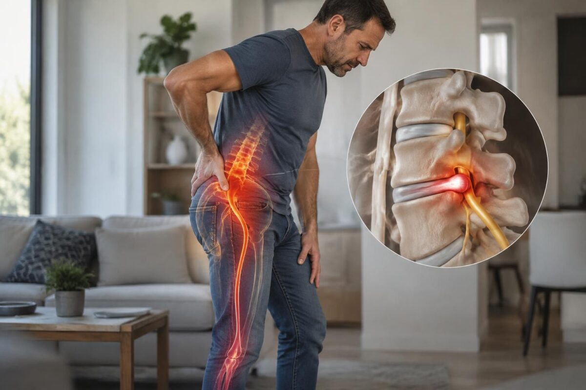
Disc herniation L4-L5 and leg pain
IN BRIEF A L4-L5 disc bulge is a protrusion of the lumbar disc that can contribute to pain in the buttock or leg if a nerve root is irritated. An MRI in a neutral position does not always show what…
Does a disc bulge always explain the pain?
No. Imaging can show slight disc changes in individuals who feel no pain. The results must align with the painful area and the signs observed during the examination. A lumbar bulge, for example, does not automatically explain pain located elsewhere.
The clinician can check if the pain, sensations, and strength follow the path of a specific nerve. They can also assess mobility and compare the results with the complete MRI report. A slight bulge without clear compression of a nerve can be an observation with no direct link to current symptoms.
Pain in the buttock can have multiple causes. It does not, therefore, confirm damage to the disc or sciatica by itself. To better grasp this subject, consult the page on disc protrusion and pain in the buttock. You can also read information on sciatica and disc protrusion.
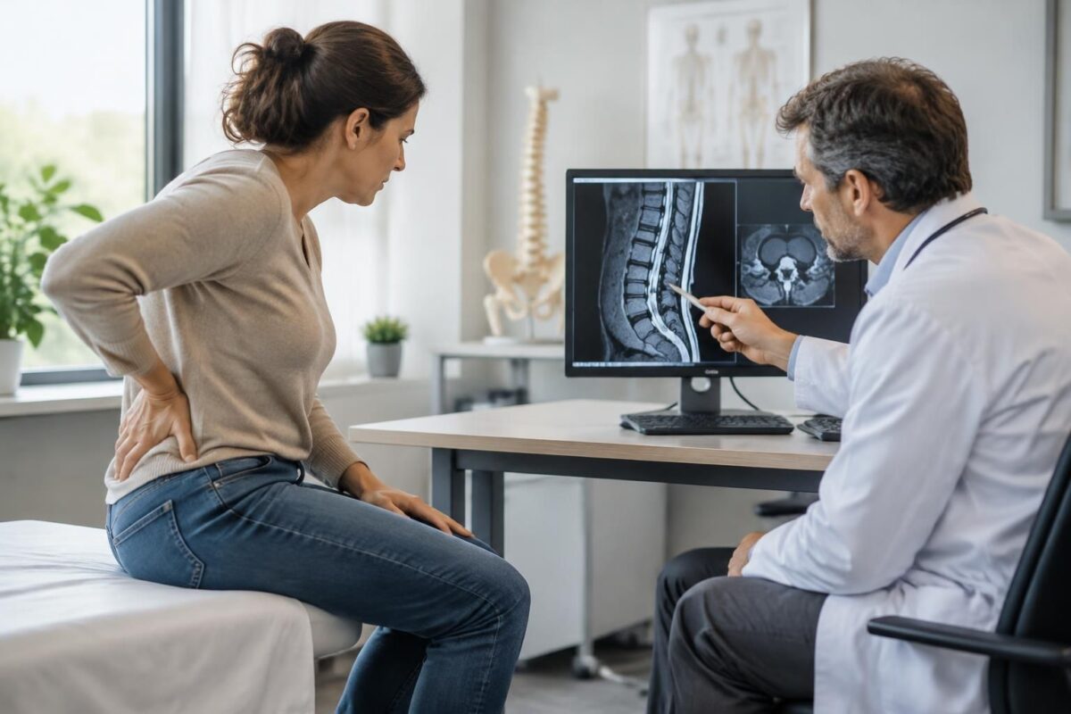
Disc bulge and sciatica: possible cause or simple discovery?
IN SHORT A disc bulge is a diffuse protrusion of a disc, sometimes associated with sciatica, but often discovered incidentally on imaging. To determine if it explains symptoms, one must compare the image to their path, the neurological examination, and…

Lumbar Disc Protrusion and the Choice of a Non-Surgical Approach
IN SHORT A disc protrusion is a bulging of a disc between the vertebrae; it often does not cause any symptoms. If it is accompanied by pain, a non-surgical approach may include appropriate activity, prescribed medications, and progressive rehabilitation. It…
How to assess a disc bulge and variable symptoms?
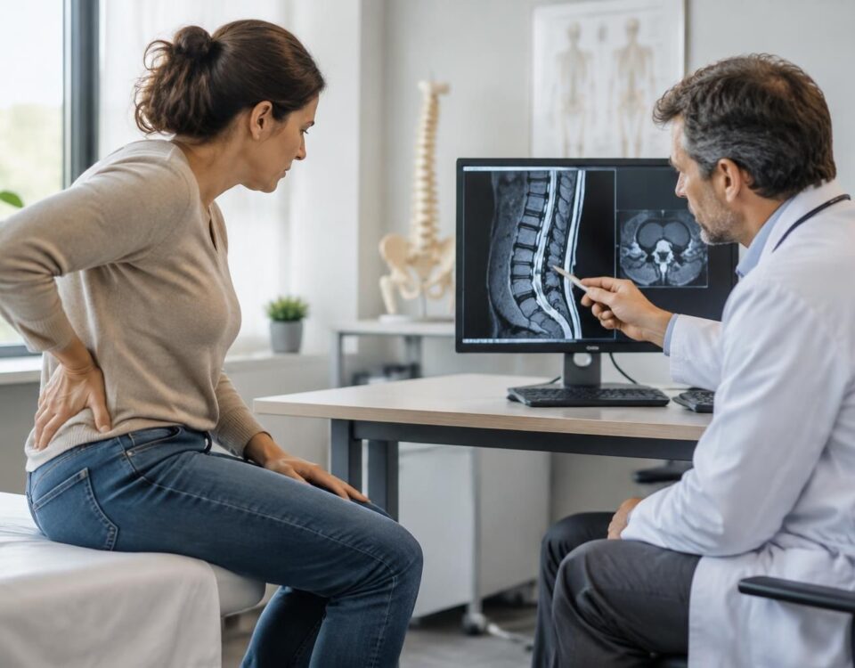
A healthcare professional takes into account the symptoms, their evolution, the patient’s history, and the results of the physical examination. They can assess strength, sensations, reflexes, and mobility. The doctor or radiologist interprets the MRI based on all these elements; the report alone does not always identify the precise cause of pain.
Depending on the situation, an MRI may show the disc, adjacent structures, and the neural exit spaces. The clinician can also examine whether the images describe a narrowing or compression that corresponds to the symptoms. To learn more about the terms used in reports, you can consult this explanation on disc bulge and disc protrusion terms only if your browser allows access to the site; however, for the resources retained here, prioritize content from SOS Sciatique and TAGMED Clinic.
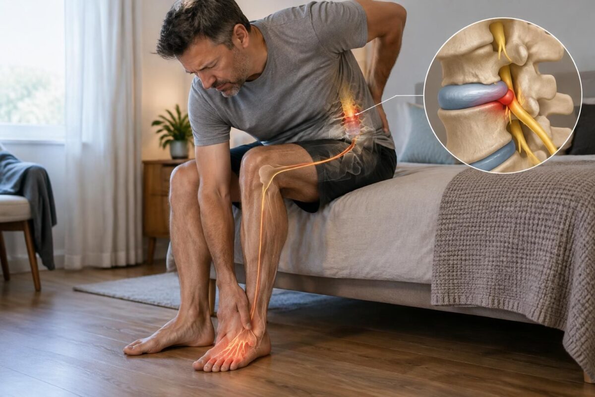
Disc herniation and pain on the top of the foot
IN BRIEF A disc protrusion is an intervertebral disc that bulges without necessarily rupturing. Pain on the top of the foot may sometimes be related to irritation of a lumbar nerve, but it does not alone confirm disc involvement. An…
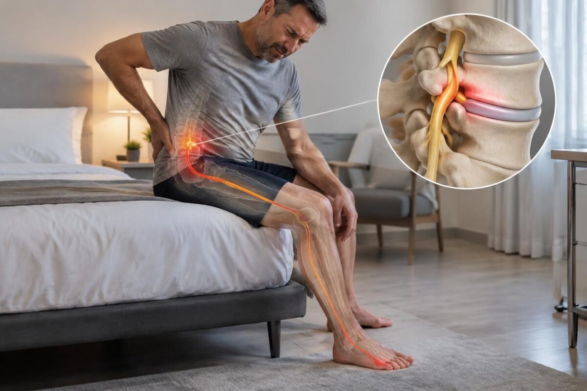
Disc protrusion and pain on the outer edge of the foot
IN BRIEF A disc bulge is a bulging of the disc that can sometimes irritate a nerve root and cause pain down to the outer edge of the foot. However, this pain is not always related to the disc: a…
What options should you discuss with a professional?
Management depends on the symptoms, examination, and goals of the person. The doctor can discuss appropriate options and determine if follow-up care or specialized advice is necessary. Surgery is not indicated solely based on the presence of a bulge on the MRI; the doctor evaluates it based on symptoms, clinical findings, and possible benefits and risks.
At the TAGMED Clinic, services may include, depending on the assessment and situation, specific osteopathy, motorized neurovertebral decompression, or precision impactor. These approaches do not replace a medical diagnosis or medical follow-up. You can check the services offered at the TAGMED Clinic in Montreal or Terrebonne, and read the general indications on consultation in case of disc protrusion.
When to quickly request an evaluation?
Quickly consult a doctor or an emergency service if you have significant or progressive weakness in a leg or an arm, new difficulty walking, loss of control over the bladder or bowels, or numbness in the saddle region. A fever associated with pain in the spine, a recent trauma, severe pain, or rapid worsening also require medical evaluation.
For persistent pain or recurring symptoms, ask a professional if the imaging results correspond to your situation. You can also inquire if a nerve root is affected and what appropriate management options are available.
About the author
Sylvain Desforges, B.Sc., D.O., N.D., Doctor of Osteopathy and naturopath, is the founding president of TAGMED and the ACMA association. He dedicates his practice to chronic pain of the spine, lower back pain, and pain compatible with sciatica, particularly when the pain radiates towards the buttock, leg, calf, foot, or toes.
Through SOS Sciatique, he publishes educational content on the symptoms of sciatica, possible causes such as disc herniation, disc pinching, foraminal stenosis, or irritation of a lumbar nerve root, as well as non-surgical approaches that may be considered based on the assessment. At the TAGMED Clinic, care may include, depending on the case, motorized neurovertebral decompression, specific osteopathy, or precision impactor.
For an evaluation related to sciatica treatment in Montreal or Mount Royal or sciatica treatment in Terrebonne, consult the services available at the TAGMED Clinic.
Medical disclaimer
The information and advice provided on this site do not replace the opinion, diagnosis, or treatment of a health professional. Please note that Sylvain Desforges, B.Sc., D.O., N.D., doctor of osteopathy, is neither a medical doctor nor a physician, and is not a specialist in a medical specialty as defined by the Collège des médecins du Québec. Manual medicine, functional medicine, and sports medicine as described on this site exclude any treatment or medical diagnosis made by a physician or medical specialist. Always consult your doctor for any medical questions. For more details, please read our full legal notice.
Why symptoms vary
How to interpret these variations

Disc protrusion and fluctuating symptoms: understanding the variations
A disc protrusion may be associated with back or neck pain, but the intensity of symptoms can vary from day to day. A painful flare-up does not necessarily mean the disc has worsened, and an MRI image is not sufficient to explain every pain. The approach to take is to observe the evolution, adapt activities without staying immobile for long, and have the results interpreted by a health professional.
Why can symptoms vary?
A protrusion corresponds to a bulging of the edge of the intervertebral disc, without necessarily rupturing its envelope. It can be visible on imaging without being the main cause of symptoms. To assess its significance, one must take into account the location, any potential nerve involvement, and the correspondence with signs observed in the clinical examination.
Pain can fluctuate depending on activities, prolonged positions, fatigue, sleep, or stress levels. Certain movements or efforts may temporarily increase sensitivity without proving that a new injury has occurred. If a nerve root is irritated, shooting pains, tingling, or numbness can also vary in intensity and sometimes spread into an arm or leg.
Observe trends rather than a single episode
Briefly note when symptoms appear, what activities influence them, and how long they last. You can also track their location and any changes in strength or sensitivity. These observations will help your clinician compare your symptoms to the MRI report, without concluding based on a single bad day.
Recommendations for managing fluctuations
Maintain movement suited to your abilities. When tolerated, alternate positions and continue daily activities at a reasonable pace. Prolonged rest can reduce physical condition and make recovery more difficult. If an activity significantly increases your symptoms, modify its intensity or duration and seek advice rather than forcing it.
Gradually resume efforts. Breaking a task into smaller parts, planning breaks, and gradually increasing activity can help better manage sensitive periods. There is no perfect posture for everyone: changing positions regularly is often more realistic than trying to remain still in an so-called ideal posture.
Have persistent or recurring symptoms evaluated. A qualified professional can assess mobility, strength, sensitivity, and reflexes, and then determine if further exams or management options should be discussed. Possible treatments depend on your situation; they may include movement support or other non-surgical care. Do not modify a prescribed treatment without consulting the responsible professional.
When to consult quickly
Request urgent evaluation for loss of bladder or bowel control, numbness in the saddle area, significant or progressive weakness in a leg or foot, or new difficulty walking. Pain accompanied by fever, occurring after significant trauma or rapidly worsening, also deserves medical attention. Beyond these signs, consult if pain persists, limits your activities, or is accompanied by new neurological symptoms.
Medical warning: this information is educational and does not replace a diagnosis or consultation. An MRI report must be interpreted by an authorized professional considering your symptoms, examination, and medical history.
| When symptoms vary | What this may mean |
|---|---|
| Stronger pain on some days | Symptoms may fluctuate according to activities and recovery. |
| Discomfort after prolonged posture | A sustained position can accentuate discomfort. |
| Pain during a specific movement | The movement may stress the back without confirming, on its own, the cause. |
| Intermittent tingling or numbness | Nerve irritation is possible; a professional can evaluate it. |
| Symptoms that change in pattern or intensity | The clinical picture must be interpreted with examination and MRI. |
| Slight bulging observed on MRI | It may be related to the symptoms or be an unrelated finding. |
| New or progressive weakness | Consult a healthcare professional quickly. |
Illustrative testimonials on disc bulge and fluctuating symptoms
Reading note: the stories below are fictitious and composite examples. They illustrate possible experiences but do not represent specific patients and do not allow for a diagnosis. A disc bulge observed on MRI must be interpreted with symptoms, clinical examination, and health history.
“My pain varied from day to day”
“My MRI report mentioned a slight lumbar disc bulge. Some days, I mainly felt stiffness in my lower back; other times, pain appeared after staying seated for a long time. The MRI reading worried me, but my healthcare provider explained that the image alone was not enough to determine the cause of my symptoms.”
“The pain sometimes radiated down my leg”
“The pain started in my lower back and sometimes extended to my buttock and thigh. It was not constant: it decreased during certain periods and then returned after more demanding days. I talked about these variations with my clinician so he could compare them to the examination and imaging results.”
“My neck and arm didn’t always hurt”
“My imaging examination described a disc bulge in my neck. My sensations changed from day to day: sometimes discomfort in the neck, sometimes tingling in the arm, and sometimes almost nothing. The evaluation allowed discussion of the location of the symptoms and to check if there were signs of nerve irritation.”
“I learned to note what influenced my symptoms”
“I noticed that discomfort varied depending on my activities, rest periods, and the duration spent in one position. Keeping notes helped me describe the changes during my consultation. This alone did not identify the cause, but it provided useful information for the assessment.”
“The report worried me less after the explanation”
“The terms ‘disc bulge’ and ‘degeneration’ made me fear a serious issue. I was explained that a slight bulge could be visible on MRI without necessarily being the main source of pain. The important thing was to verify whether its location matched my symptoms and if there was significant nerve compression.”
Fluctuating symptoms can be influenced by several factors, including activities, prolonged postures, sleep, and overall health status. Their evolution alone does not confirm or exclude a link to a disc bulge. To understand the significance of an MRI result, discuss it with a qualified healthcare professional.
Consult a doctor or emergency service quickly in case of loss of bladder or bowel control, numbness in the saddle area, significant or progressive weakness in a leg or foot, difficulty walking, fever associated with back pain, recent trauma, or rapid worsening.
Disc bulge and fluctuating symptoms: understanding the variations
A disc bulge describes an intervertebral disc whose contour slightly exceeds its usual limit, without necessarily presenting a rupture. Discs, located between vertebrae, help absorb stress and allow movement of the spine. A bulge can be observed in the neck or lower back, but its presence on imaging does not automatically mean it is the cause of the pain felt.
Associated symptoms may be fluctuating: they vary in intensity, appear in episodes, or change according to activities and positions. A person may feel moderate discomfort on some days, then more pronounced pain after a prolonged sitting period, unusual effort, or a difficult night of sleep. This variation alone does not conclude that the disc has worsened. Tissues, muscles, joints, and the nervous system can react differently depending on the circumstances.
The location of the bulge and its relation to neighboring structures matter more than just the term recorded in a report. If a nerve root is irritated, pain can sometimes radiate from the neck to the shoulder, arm, or hand, or from the lower back to the buttock and leg. Symptoms such as tingling, numbness, or weakness may also occur. However, these manifestations have several possible causes and require appropriate evaluation.
Symptoms can change with movements, the duration spent in a posture, the level of activity, or recovery. Stress, sleep, and overall health status can also influence pain perception. Therefore, a painful flare-up does not necessarily mean there is a new injury. Conversely, persistent or progressively worsening symptoms warrant discussion with a healthcare professional.
An MRI result must be interpreted in light of symptoms, physical examination, and history. Mild bulges and age-related changes are common, including in individuals who report no pain. The clinician seeks to determine if the level described in imaging corresponds to the painful area and whether there is any narrowing of the canal or openings where nerves pass. Imaging is part of the evaluation, not a diagnosis in itself.
Management depends on the individual situation. After an evaluation, non-surgical options can be discussed with a qualified professional. These may include advice on activity, tailored exercises, physiotherapy, or other appropriate approaches. The goal is generally to enhance function and better manage symptoms, without assuming that one method fits all. Surgical intervention is only considered in certain cases, according to findings, progression, and the person’s goals.
Seek medical attention promptly if there is a new or progressive weakness, difficulty walking, numbness in the saddle area, or loss of bladder or bowel control. A fever accompanied by spinal pain, significant trauma, or rapid deterioration also requires medical evaluation.
About the author
Sylvain Desforges, B.Sc., D.O., N.D., Doctor of Osteopathy and naturopathy, is the founding president of TAGMED clinics and the ACMA association. His practice focuses in particular on chronic spinal pain and functional support. Depending on the evaluation and situation, the care offered in the clinic may include specific osteopathy, motorized neurovertebral decompression, or precision percussion.
Medical Disclaimer
This content is provided for educational purposes and does not replace a consultation, diagnosis, or treatment by a physician or other authorized healthcare professional. The author is neither a physician nor a specialist in a medical specialty recognized by the Collège des médecins du Québec. Approaches such as osteopathy, manual medicine, or functional support do not constitute medical acts and do not replace medical follow-up.
A bulging disc may be associated with pain that varies from day to day, but its presence on an MRI is not sufficient to explain every symptom. The significance of this finding depends in part on its location, its potential effect on the nerves, and its correspondence with the pain, numbness, or weakness felt. A clinical evaluation helps to place the images in context.
Why can symptoms fluctuate?
Pain can change according to activities, positions, the duration spent sitting, sleep, fatigue level, or repeated strains. Certain movements may temporarily worsen symptoms; other moments of rest or appropriate activity may alleviate them. These variations alone do not determine whether a disc is involved or if the situation is worsening. For example, low back pain in a seated position may have several explanations, even when a disc cause is considered: sciatic pain worsened in sitting position.
A bulge corresponds to a protrusion of the disc’s contour; it does not automatically mean that a nerve is compressed. To better understand what the term “bulging” encompasses, please refer to this explanation of disc bulging. Symptoms may also arise from other structures of the back or neck. Therefore, a bulging disc may be a possible cause, an associated finding, or an element unrelated to the current pain: bulging disc and sciatica: possible cause or incidental finding.
Linking the MRI to the symptoms
The healthcare professional relates the imaging report to the history of symptoms and the physical examination. Pain that radiates down to the buttock or leg, tingling, loss of sensation, or weakness may guide the evaluation towards nerve irritation, but none of these signs alone confirms a diagnosis. The specifics of lumbar disc involvement with radiation are addressed in this article on sciatica and lumbar disc herniation.
Vertebral level also matters: involvement at L4-L5 can be examined based on the location of symptoms and neurological findings. To learn more about manifestations that may be associated with this level, consult the page dedicated to sciatica L4-L5. The terms in the report and possible symptoms of the bulge are also presented by Clinique Solutions Discale along with their page on symptoms associated with disc bulging.
At the cervical level, a bulge may be examined more closely if symptoms appear in the shoulder, arm, or hand. The presentation of cervical disc bulging can help understand possible symptoms, without replacing personalized interpretation from an examination. General information about bulging discs is also available from Continental Hospitals.
Adapting activities and requesting an evaluation
When symptoms fluctuate, it is generally helpful to note the activities, positions, and circumstances that modify them, and then discuss them with a qualified professional. Avoiding any activity for fear of an MRI result is not always necessary; conversely, it may be wise to temporarily adjust a movement that clearly triggers pain. Lumbar flexion and disc symptoms are addressed in this article on caution during lumbar flexion. Management options should take into account the examination, history, and evolution of symptoms.
Consult a doctor or another licensed healthcare professional if pain persists, worsens, or limits daily activities. An urgent evaluation is necessary in case of loss of bowel or bladder control, numbness in the saddle region, significant or progressive weakness, new difficulty walking, fever with spinal pain, or recent trauma. The information presented here is educational and does not replace a diagnosis or medical advice.

Sylvain Desforges, B.Sc., D.O., N.D., docteur en ostéopathie
Dr Sylvain Desforges, B.Sc., D.O., N.D., osteopath, is the founding president of TAGMED and of the ACMA association. He dedicates his practice to chronic spinal pain, low back pain and symptoms compatible with sciatica, particularly when pain radiates toward the buttock, leg, calf, foot or toes.
Through SOS Sciatique, he publishes educational content on sciatica symptoms, possible causes such as disc herniation, disc narrowing, foraminal stenosis or irritation of a lumbar nerve root, as well as non-surgical approaches that may be considered according to the evaluation. At Clinique TAGMED, care may include, depending on the case, motorized neurovertebral decompression, specific osteopathic care or precision percussion.
For an evaluation related to sciatica treatment in Montreal / Mount Royal or sciatica treatment in Terrebonne, please consult the services available at Clinique TAGMED.
Medical disclaimer
The information published on SOS Sciatique is provided for educational purposes only and does not replace a consultation, diagnosis, opinion or treatment provided by a physician or another authorized healthcare professional. The author of this article is neither a medical doctor nor a specialist in a medical specialty recognized by the Collège des médecins du Québec. The osteopathic, manual medicine or functional support approaches described on this site do not constitute medical acts and are not intended to replace medical follow-up.
Consult a physician or emergency service in cases of loss of bladder or bowel control, anesthesia in the saddle area, significant or progressive weakness in the leg or foot, fever, recent trauma, severe pain or rapid worsening of symptoms. For more details, please read our Legal Notice.