



Sciatica and pain on one side: possible causes
14/08/2026
Sciatica and foot pain in the car
14/08/2026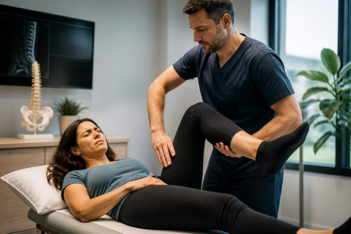
IN BRIEF
In brief, the sequestered disc herniation and sciatica describe a root compression due to a detached disc fragment that follows the typical pathway of sciatica. The evaluation is crucial: it confirms the diagnosis, specifies the affected level (L4-L5 or L5-S1), detects any potential neurological emergency, and guides treatment between conservative options and surgical interventions. With the help of MRI and clinical examination, the risk of motor deficit can be estimated, rehabilitation can be planned, and recovery can be accelerated. Accessible in Montreal, this early evaluation improves recovery chances and avoids therapeutic delays. |
The sequestered disc herniation is an advanced form of sciatica where a fragment of the disc nucleus detaches and can severely compress one or more nerve roots. It is often accompanied by intense pain, paresthesias, and potential motor deficits.
The evaluation is essential to confirm the diagnosis, determine the level and type of herniation (e.g., L4-L5 or L5-S1), and detect warning signs that require prompt management.
It is based on a thorough clinical examination and a lumbar MRI, a key examination before any therapeutic decision. This evaluation guides the choice between conservative treatment (relative rest, anti-inflammatories, physiotherapy, injections if necessary) and surgical or minimally invasive intervention when motor deficit is present or when pain persists.
In summary, a quick and comprehensive evaluation enhances safety, accelerates recovery, and helps make the best therapeutic decisions.
Quick summary: This article explains what a sequestered disc herniation is and why the evaluation is essential in the face of sciatica. The evaluation combines a precise clinical examination and appropriate imaging to identify the level and type of herniation, assess risks, and guide decision-making, particularly in cases of neurological deficit or persistent pain. It allows for distinguishing situations requiring conservative treatment from those needing intervention and anticipating outcomes, including when surgery may be considered.
What is a sequestered disc herniation?
A sequestered disc herniation occurs when a fragment of the nucleus pulposus detaches from the disc and moves freely in the spinal canal. This fragment can compress one or more nerve roots. In the context of sciatica, the location and course of pain depend on the affected level. A sequestration can cause intense radicular pain and neurological disturbances if the compression is significant.
How is the evaluation conducted?
The evaluation relies on two pillars: clinical examination and imaging. The clinical examination looks for signs of radiculopathy, loss of strength, or sensory disturbances in the corresponding territory. Tests like the Lasègue sign, reflex assessment, and strength of the foot dorsiflexors help clarify the affected level and severity of symptoms. The lumbar MRI is the reference examination to confirm the presence of a sequestered disc herniation, specify its level (e.g., L4-L5 or L5-S1), and assess the extent of nerve compression. The evaluation also helps eliminate other causes of low back and radicular pain.
Role of imaging and diagnosis
The lumbar MRI identifies the type of herniation (protrusion, extrusion, or sequestration) and specifies the exact level. It assesses the degree of nerve root compression and helps plan treatment. The MRI should be performed on a suitable machine and focused on the affected levels. It is not systematically done at the beginning when symptoms are recent and mild but becomes essential when symptoms persist or worsen, or in the presence of neurological signs.
When does the evaluation lead to an intervention?
The prognosis is favorable in the majority of cases when the evaluation leads to conservative treatment. An intervention becomes feasible in cases of progressive motor deficit, such as the onset of a foot drop, or when the pain is hyperalgesic and debilitating despite optimal medical treatment for several weeks. The presence of a cauda equina syndrome or loss of control of urinary or bowel functions is an absolute emergency requiring prompt intervention. A precise evaluation distinguishes cases where surgery may offer lasting relief from those where conservative treatment is preferred.
Resources and further readings
To delve deeper, please refer to the following resources:
- Herniated disc and sciatica – general resources
- Information on sequestered disc herniation and management options
- Extruded disc herniation and sciatica – clinical significance
- TAGMED Clinic – Information and resources
- Neurovertebral decompression – concepts and indications
- Sciatica and heel pain – possible S1 root
- Professional resources – osteopathy and associated pathologies
For more information on mechanisms and management options, additional readings and reliable clinical resources can be consulted on the above sites.
Medical disclaimer: The information and advice provided on this site do not replace the opinion, diagnosis, or treatment of a healthcare professional. Please note that Dr. Sylvain Desforges, osteopath, is neither a doctor of medicine nor a physician, and is not a specialist in a medical specialty as defined by the Collège des médecins du Québec. Manual therapy, functional medicine, and sports medicine as described on this site exclude any medical treatment or diagnosis made by a physician or a specialist doctor. Always consult your doctor for any medical questions. For more details, please read our complete Legal Notice.
- Assessment Axis: determine if the sequestration is the main cause and its impact on the nerve to guide therapeutic decision-making.
- Clinical Signs: radicular pain, weakness or paresthesia in the L5/S1 territory; presence of foot drop or cruralgia.
- Neurological Urgency: progressive motor deficit or signs of cauda equina requiring prompt consultation.
- Functional Impact: walking ability and daily activities that influence the choice of management.
- Imaging and Diagnosis: lumbar MRI specifies the type (sequestration/extrusion/protrusion) and the exact level.
- Therapeutic Plan: the evaluation guides between conservative and surgery.
- Preparation and Safety: assessment of comorbidities and organization of pre/post-operative rehabilitation.
- Surgical Techniques: choice between microdiscectomy and endoscopy based on anatomy and patient preferences.

This article presents why evaluation is essential in case of sequestered disc herniation associated with sciatica. It describes what sequestration is, the warning signs, the role of imaging, and how evaluation guides therapeutic choices, with an emphasis on the speed and accuracy needed to optimize prognosis.
What is a sequestered disc herniation and why does sciatica occur
A sequestered disc herniation corresponds to a fragment of the nucleus pulposus that detaches from the disc and migrates into the spinal canal. This migration can exert greater pressure on a nerve root, worsening pain and neuro-sensitivity along the path of the affected nerve. Understanding this mechanism helps anticipate the evolution of symptoms and distinguish this pathology from other causes of low back pain or radiculopathy.
The associated sciatica can then follow typical pathways depending on the level affected. Precise evaluation allows for identifying not only the location of the compression but also any emerging motor deficit, which will guide early management and the timing of treatments.
Key elements of the evaluation
Clinical Examination
During the examination, the doctor looks for specific signs such as reproduction of radicular pain through simple tests, the Lasègue sign, and assessment of muscle strength (foot dorsiflexors, quadriceps, plantar flexors). Osteotendinous reflexes and sensitivity in the radicular territories help localize the affected root (L5 or S1 and beyond depending on the level). The presence of a dropped foot or progressive weakness may signal a relative urgency and require prompt evaluation by a specialist.
Role of Imaging
The MRI of the lumbar spine is the key tool in diagnosis. It confirms the presence of a herniation, specifies the exact level (e.g., L4-L5 or L5-S1), describes the type of herniation (protrusion, extrusion, sequestration), and evaluates the degree of nerve compression. Imaging complements the clinical examination and allows for planning the most appropriate treatment, while looking for any other potential causes of pain.
Warning Signs and Urgency
Certain elements require prompt management: marked or recent motor deficit (foot drop), very intense pain that does not subside despite treatment, walking disturbances, or signs indicating a risk to the roots. In these cases, a quick consultation with a neurosurgeon is recommended to discuss the potential for intervention and the optimal timing.
Why evaluation influences therapeutic choices
A precise evaluation allows for distinguishing situations that may benefit from conservative treatment (medications, targeted physiotherapy, injections) from those that require rapid surgical intervention, particularly in the presence of sequestration associated with nerve compression. Timing is crucial: when symptoms reflect significant nerve pressure and a motor deficit exists or progresses, the surgical option may offer a more favorable recovery, especially if the pain is disabling and persists beyond non-surgical attempts.
Furthermore, the evaluation includes planning for post-treatment rehabilitation and muscle strengthening, in order to prevent recurrences and optimize the stability of the lumbar spine. A personalized approach, adapted to the location of the herniation and the level of involvement, improves the chances of functional recovery.
| Key Aspect | Concise Elements |
|---|---|
| Initial Clinical Signs | Identify the criteria that guide the evaluation and choice of treatment: typical radicular pain and irradiation, with signs such as Lasègue and sensory disturbances. |
| Motor Deficits | Monitor foot drop and strength of foot dorsiflexors; any progressive deficit warrants prompt consultation. |
| Neurological Emergencies | Cauda equina syndrome and rapid weakness or loss of bladder control require prompt evaluation to avoid complications. |
| Role of Imaging | Lumbosacral MRI confirms the level and type of herniation, evaluates compression, and guides therapeutic decisions. |
| Initial Treatment | The conservative treatment aims to relieve pain and promote resorption: relative rest, NSAIDs, analgesics, and progressive physiotherapy after the acute phase. |
| Imaging Limitations | Imaging may not be indicated in very recent phases without signs of severity; MRI complements the clinical evaluation depending on the context. |
| Surgical Indications | Surgery considered after failure of conservative treatment (around 6-8 weeks) or in the presence of clearly established neurological emergencies. |
| Decompression Techniques | Microsurgical or endoscopic discectomy to relieve the root; choice adapted to the anatomy and preferences of the patient. |
| Follow-up and Prevention | Lumbar strengthening, weight control, smoking cessation, and gradual resumption of activities to limit recurrences. |
Testimonials on the Evaluation of Sequestered Disc Herniation and Sciatica: Why Evaluation is Important
Testimonial 1: Before my first consultation, I thought it was simply lower back pain that would go away with rest. Thanks to a complete evaluation, a sequestered disc herniation at the L5-S1 level was identified, compressing the S1 root. The combination of clinical examination and MRI clarified the diagnosis and guided treatment, avoiding delays and complications.
Testimonial 2: My symptoms seemed to follow the usual pain patterns, but the evaluation allowed for precise differentiation of painful areas and identified a sciatica related to a key location. Understanding that each level can cause different symptoms, depending on the affected root, gave me confidence in the choice of therapeutic pathway and in preventing future exacerbations.
Testimonial 3: After several weeks of pain and numbness, the evaluation revealed a femoral nerve pain associated with a disc herniation at L3-L4. Knowing exactly which root was affected allowed me to understand why the pain radiated down the front of my thigh and why certain movements were more painful than others, guiding management without unnecessary delays.
Testimonial 4: When I began to show foot drop, rapid evaluation was crucial. The doctor assessed the motor deficit and planned an appropriate intervention within a critical timeframe, which can influence recovery and limit long-term sequelae.
Testimonial 5: The evaluation also helped determine the precise type of disc herniation, whether protrusive, extrusive, or sequestered, as well as the exact level (L4-L5 or L5-S1). This precision is essential for selecting the most suitable therapeutic plan and anticipating subsequent steps, such as non-surgical or surgical options.
Testimonial 6: Finally, early evaluation shows that most episodes resolve without surgery, but careful follow-up and appropriate rehabilitation, guided by reliable evaluation, help limit pain and promote a rapid and sustainable recovery of walking and daily activities.
Evaluation of Sequestered Disc Herniation and Sciatica: Why Evaluation is Crucial
Dr. Sylvain Desforges, an osteopath with expertise in a holistic approach to pain and manual therapies, has made it his mission to help patients understand the importance of a rigorous evaluation in the face of a sequestered disc herniation and sciatica. With experience in integrating advanced technologies — spinal decompression, laser, and shockwave therapy — his practice is based on precise evaluation that guides every step of the care pathway toward effective and sustainable recovery.
The disc sequestration represents a particular stage of disc pathology where a fragment of the nucleus pulposus detaches and migrates into the spinal canal. This migration can cause more pronounced nerve compression and sometimes extremely intense pain, with an increased risk of evolving into specific neurological symptoms. Thorough evaluation not only confirms the diagnosis but also distinguishes this situation from other causes of low back and radicular pain, preventing therapeutic delays that could compromise nerve function.
A detailed clinical evaluation is the foundation of management. The interview focuses on describing the painful trajectory, irradiated areas, and motor-sensory signs. The clinician looks for revealing clinical elements, such as signs of Lasègue, integrity of reflexes (Achilles and patellar), and strength of foot dorsiflexors, plantar flexors, and quadriceps. In the case of femoral nerve pain or a major motor deficit, the evaluation must quickly clarify the potential urgency for specialized management to prevent lasting sequelae.
The role of imaging is crucial. Lumbosacral MRI is the reference examination, allowing for the identification of the precise level (often L4-L5 or L5-S1), characterization of the type of herniation (protrusion, extrusion, or sequestration), and estimation of the degree of nerve compression. This step is essential for planning any potential surgical intervention or adapting the non-surgical therapeutic plan. An appropriate radiological evaluation, combined with rigorous clinical analysis, helps avoid misguided treatments and optimizes recovery chances.
Care is not uniformly surgical. Most patients show improvement with conservative measures and appropriate rehabilitation. However, when the assessment reveals significant nerve compression or progressive motor deficit, intervention may be necessary and beneficial if performed early. The evaluation serves as a compass to distinguish situations where a delay may be tolerable from a case where a rapid approach is indicated to preserve motor function and quality of life.
At the heart of his approach, Dr Sylvain Desforges emphasizes a global coordination of care. Beyond osteopathic care and manual techniques, he integrates modern tools and a patient-centered approach to optimize well-being and recovery. His goal is to propose evidence-based strategies while remaining attentive to the individual needs of each patient and providing support that encourages a gradual return to daily activity, followed by thoughtful and adapted sports practice.
About the author
Dr Sylvain Desforges, B.Sc., D.O., N.D., osteopath, is the founding president of
TAGMED and the ACMA association. He dedicates his practice to chronic pain of the spine, lower back pain, and pain compatible with sciatica, particularly when the pain radiates to the buttock, leg, calf, foot, or toes.
Through SOS Sciatique, he publishes educational content on sciatic symptoms, possible causes such as herniated disc, disc pinching, foraminal stenosis, or irritation of a lumbar nerve root, as well as the non-surgical approaches that may be considered based on the assessment. At the TAGMED Clinic, care may include, depending on the case, motorized neurovertebral decompression, specific osteopathy, or precision percussion.
For an evaluation related to
sciatica treatment in Montreal / Mont-Royal
or to
sciatica treatment in Terrebonne,
please refer to the services available at the
TAGMED Clinic.
Medical disclaimer
The information published on SOS Sciatique is provided for educational purposes only and does not replace a consultation, diagnosis, advice, or treatment by a physician or another authorized health professional. The author of this article is neither a physician nor a specialist in a medical condition recognized by the Collège des médecins du Québec. The approaches of osteopathy, manual medicine, or functional support described on this site do not constitute medical acts and are not intended to replace medical follow-up.
Consult a physician or emergency service in case of loss of bladder or bowel control, anesthesia in the saddle area, significant or progressive weakness of the leg or foot, fever, recent trauma, severe pain, or rapid worsening. For more details, please read our
Legal Notice.
In summary, this article reminds us that evaluation is the pivot of managing sequestrated disc herniation associated with sciatica. A good analysis allows distinguishing levels L4-L5 and L5-S1, assessing neurological signs, and deciding whether a conservative approach is sufficient or if surgical decompression is necessary.
A comprehensive evaluation is the foundation of managing sequestrated disc herniation with sciatica. It combines a precise history of symptoms and a targeted clinical examination (Lasègue’s sign, strength of foot dorsiflexors, reflexes, sensitivity) with appropriate imaging to confirm the level and type of hernia (protrusion, extrusion, or sequestration).
A precise diagnosis not only guides the choice between conservative treatment and intervention but also the speed of intervention in case of nerve compromise. The onset of a foot drop or progressive motor deficit constitutes a relative emergency requiring a rapid neurosurgical consultation (generally within 24 to 72 hours). Such a recent deficit is likely to recover after surgery if the intervention is performed early.
The anatomo-clinical aspects of levels L4-L5 and L5-S1 explain why distinct symptoms appear depending on the affected root. Disc sequestration can cause intense pain and marked neurological discomfort, requiring thorough assessment to rule out serious causes and plan an appropriate therapeutic strategy. Specialized resources and clinical guidelines emphasize the importance of accurate diagnosis and rigorous follow-up to optimize outcomes.
For better understanding and practical perspectives on mechanisms, decompression options, and non-surgical approaches, you can consult complementary resources such as Doctissimo, Clinique duDos, or SOS Sciatique. Other specialized resources remind us of the importance of a comprehensive evaluation before any surgical decision and present the potential benefits of neurovertebral decompression and minimally invasive options, such as lumbar endoscopy, when the indications are met.
A early and comprehensive evaluation thus helps avoid delays in diagnosis, prevent irreversible motor deficits, and direct towards the most appropriate therapeutic strategy, whether non-surgical or surgical. For patients showing signs of severity or not responding to appropriate conservative treatment, a rapid specialized consultation can make the difference between quick recovery and lasting sequelae.
To explore diagnostic and treatment options in depth, see in particular the specialized resources in spine surgery and the management guides provided by dedicated teams in Paris and online: Lumbar disc herniation L4-L5, Decompression without surgery, and gentle and effective approach.

Disc herniation and associated foraminal stenosis
IN BRIEF A disc protrusion associated with foraminal stenosis occurs when a bulging disc coexists with a narrowing of the opening through which a nerve root passes. This association can lead to lower back pain radiating into the leg, tingling,…

Spinal disc protrusion and nerve inflammation
IN BRIEF A disc protrusion is a bulging of the disc that can irritate or compress a nerve, without necessarily causing symptoms. When it leads to nerve inflammation, it can cause pain in the back or neck, sometimes accompanied by…

Disc protrusion and irritation of L5
IN SHORT A disc protrusion is a bulging of the disc that can irritate the L5 root and cause lower back pain radiating to the buttock, leg, or foot, sometimes with tingling or weakness. MRI alone is not sufficient to…

Disc protrusion and pain after twisting the back
IN BRIEF A disc protrusion is a bulging of the disc that can contribute to pain after a back twist, but it alone does not confirm the cause. The pain can remain in the lower back or radiate to the…

Disc herniation and pain after lifting
IN BRIEF A disc protrusion is a bulging of a disc between the vertebrae. Pain that appears after lifting a load does not prove that it is the cause: the MRI must be interpreted alongside the symptoms and the clinical…

Disc herniation and lumbar flexion: why caution is necessary
IN BRIEF A disc protrusion is a bulge of the intervertebral disc, which can be silent or irritate a nerve. Lumbar flexion is not necessarily dangerous but can worsen symptoms in some individuals. Adapting movements, avoiding those that aggravate pain,…

Disc herniation and pain that worsens in the car
IN BRIEF A disc protrusion is a bulging of a disc that can irritate a nerve. In a car, prolonged sitting and vibrations can exacerbate back, buttock, leg, or arm pain. Regular breaks, an adapted posture, and personalized management can…

Disc protrusion and pain that increases when sitting
IN BRIEF A disc protrusion is an intervertebral disc that bulges without necessarily breaking its outer covering. Pain may increase when sitting and sometimes radiate into the buttock or leg, like sciatica. Alternating positions, moving regularly, and following a suitable…

Dr Sylvain Desforges, B.Sc., D.O., N.D., osteopath, is the founding president of TAGMED and of the ACMA association. He dedicates his practice to chronic spinal pain, low back pain and symptoms compatible with sciatica, particularly when pain radiates toward the buttock, leg, calf, foot or toes.
Through SOS Sciatique, he publishes educational content on sciatica symptoms, possible causes such as disc herniation, disc narrowing, foraminal stenosis or irritation of a lumbar nerve root, as well as non-surgical approaches that may be considered according to the evaluation. At Clinique TAGMED, care may include, depending on the case, motorized neurovertebral decompression, specific osteopathic care or precision percussion.
For an evaluation related to sciatica treatment in Montreal / Mount Royal or sciatica treatment in Terrebonne, please consult the services available at Clinique TAGMED.
Medical disclaimer
The information published on SOS Sciatique is provided for educational purposes only and does not replace a consultation, diagnosis, opinion or treatment provided by a physician or another authorized healthcare professional. The author of this article is neither a medical doctor nor a specialist in a medical specialty recognized by the Collège des médecins du Québec. The osteopathic, manual medicine or functional support approaches described on this site do not constitute medical acts and are not intended to replace medical follow-up.
Consult a physician or emergency service in cases of loss of bladder or bowel control, anesthesia in the saddle area, significant or progressive weakness in the leg or foot, fever, recent trauma, severe pain or rapid worsening of symptoms. For more details, please read our Legal Notice.