


Sciatica and pain when laughing or straining: possible disc pressure
26/09/2026
Sciatica and Insoles: What You Need to Understand Without Exaggeration
26/09/2026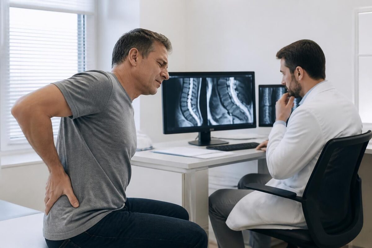
IN BRIEF In brief, Herniated disc and sciatica are not determined by MRI alone. The diagnosis relies on clinical coherence between the symptoms and physical examination, which guides the subsequent steps of monitoring, treatment, and targeted imaging. Advantages include avoiding unnecessary imaging, directing towards a gradual treatment and rehabilitation, and reducing fear and delays in care. Functioning: a comprehensive clinical evaluation analyzes the painful trajectory and neurological signs; MRI becomes useful when pain persists or when specific localization is needed before making a surgical decision or infiltration. Available in Montreal, this service is particularly useful to respond quickly to an evaluation need without over-imaging and to prioritize a global and gradual approach. |
The diagnosis of herniated disc and sciatica is primarily based on the coherence between symptoms and clinical examination, rather than on imaging alone. The MRI can show the discs, roots, and spinal canal, but it does not evaluate the functional nerve irritation, sensitivity, or the impact of movement on the nerve. A visible herniated disc is not necessarily painful, and a small hernia can cause significant pain depending on orientation and root irritation.
The diagnosis is based on the trajectory of the pain, neurological signs (weakness, numbness, loss of strength), and clinical tests, not on imaging alone. MRI is useful in cases of persistent pain, diagnostic uncertainty, or before an interventional procedure, but it does not determine the therapeutic approach by itself. In practice, a gradual approach centered on movement and function is preferred; surgery is considered only after a comprehensive evaluation and according to the patient’s progress.
Herniated disc and sciatica: what the MRI alone cannot decide explains why imaging, even very detailed, does not solely determine treatment. Effective diagnosis relies on the coherence between symptoms, clinical examination, and the overall assessment of the patient. This article guides you to understand what the MRI can and cannot say, and how a progressive and adapted approach is built from all clinical elements.
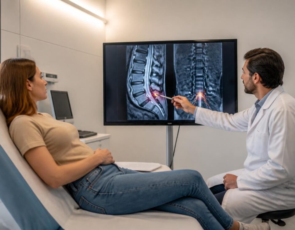
The MRI provides accurate images of the spinal structures, but it does not make the pain visible and does not indicate whether a hernia is truly responsible for the symptoms. A herniated disc may appear on imaging in people without pain, while conversely, strong pain may exist without an identifiable hernia. This is why the diagnosis primarily relies on clinical evaluation and the correspondence between signs and symptoms.
The diagnosis relies on symptomatic and clinical coherence
The physician analyzes the trajectory of the pain. Pain limited to the lower back may reflect a lumbago or discopathy without evident nerve compression. If the pain descends into the buttock, back of the thigh, calf, or foot, sciatica is more likely considered. Pain traveling toward the front of the thigh or knee suggests cruralgia. A precise description of the trajectory helps identify the potentially compressed nerve root.
The role of the clinical examination
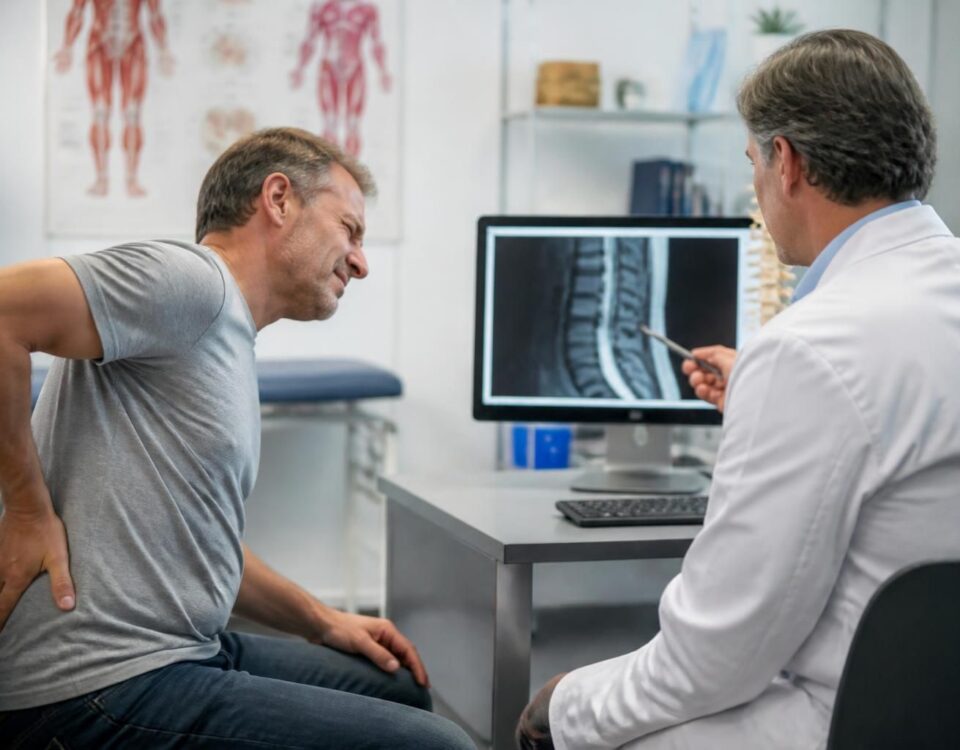
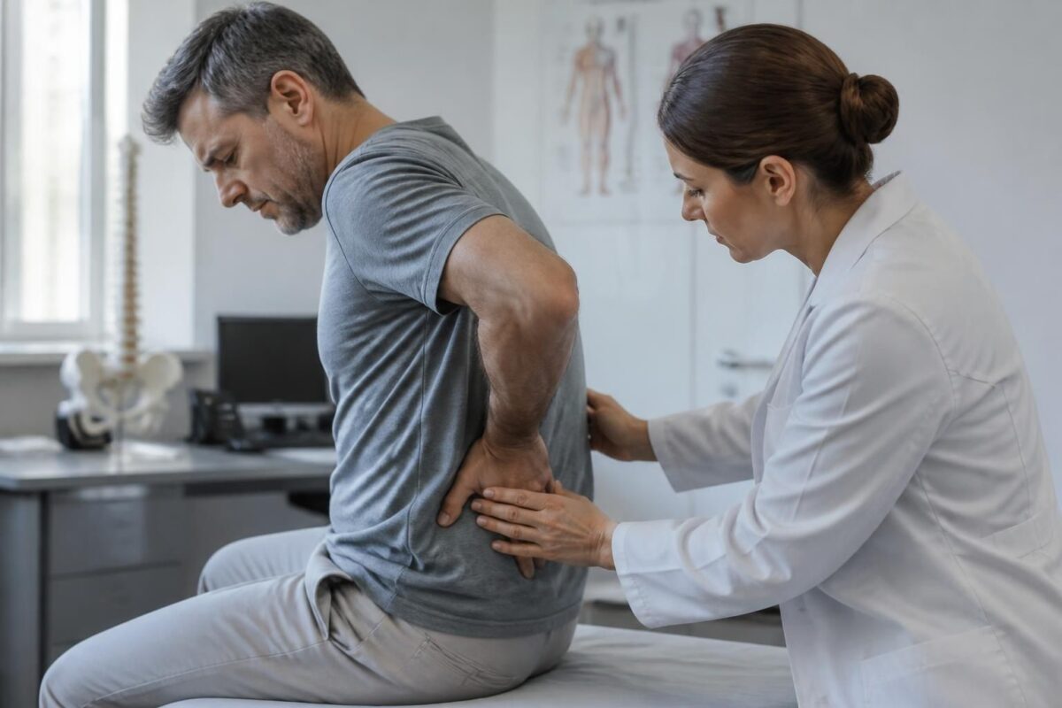
The clinical examination is based on the interview and physical tests. The mobility of the lumbar spine, pain during certain movements, reflexes, muscle strength, and sensitivity are assessed. Nerve tension tests may reproduce pain in the leg and guide the diagnosis. The clinical examination helps avoid classic errors: attributing all back pain to a herniated disc or becoming alarmed by an image that does not correspond to the symptoms.
When MRI is useful and when it may be unnecessary
The MRI becomes useful when pain persists despite consistent management, when there is a neurological deficit, or when a targeted intervention is considered. In most cases, lumbar pain improves naturally within a few weeks, and a herniated disc spontaneously resolves in the vast majority of situations. In this context, imaging does not necessarily accelerate healing and should not replace clinical reasoning.
What MRI shows and what it does not
The MRI shows the discs, vertebrae, nerve roots, and soft tissues of the spinal canal. However, it does not measure functional nerve irritation, the sensitivity of the nervous system, the stress associated with pain, or the overall impact of movements on the patient. An image is not an explanation; it remains just one piece of information among other elements to gather for diagnosis.
Integration of complementary examinations
Complementary examinations must be chosen based on clinical evolution. MRI is particularly useful in the presence of a progressive neurological deficit, unexplained pain, or preoperative assessment. CT scans may be considered when MRI is not possible or according to medical indications. X-rays provide information on alignment and arthritis but do not directly show the hernia.
To enrich your understanding, you can consult resources that explain when imaging is useful and when it may complicate the diagnostic process, notably Sciatica without herniated disc and Herniated disc and pain down the leg.
Reevaluation and progressive management
A progressive approach prioritizes movement, reducing fear, and restoring suitable activities. Rehabilitation, posture advice, and improving daily movements play a key role. When pain persists despite targeted management, additional options may be discussed with the patient and relevant specialists.
Complementary resources
To deepen your understanding of concepts related to neurovertebral decompression and the management of lumbar pain, you can consult the following resources:
Herniated disc and mechanical nerve compression
Sequestered herniated disc and sciatica
Herniated disc and chronic pain
Sciatica without herniated disc
For management options in Montreal or Terrebonne, you can turn to TAGMED Clinic.
For additional information on imaging and sciatica, you can also consult the following resources:
To delve deeper and compare approaches, associated resources help clarify choices between imaging and clinical management, notably through the links provided above.
Medical disclaimer: The information and advice provided on this site do not substitute for the opinion, diagnosis, or treatment of a healthcare professional. Please note that Dr. Sylvain Desforges, osteopath, is neither a medical doctor nor a physician, and is not a specialist in a medical specialty as defined by the Collège des médecins du Québec. Manual medicine, functional medicine, and sports medicine as described on this site exclude any treatment or medical diagnosis performed by a physician or specialist. Always consult your doctor for any medical questions. For more details, please read our complete Legal Disclaimer.
- The clinical correlation: pain and neurological signs are not limited to the image.
- The clinical diagnosis: symptoms + examination remain essential.
- The surgical decision: an image does not decide; conservative treatment is often sufficient.
- The prognosis: healing possible without intervention; imaging does not predict it.
- The therapeutic plan: rehabilitation, medication, and injections depend on the clinical course.
- Exact location: MRI helps locate the compression but must be validated by symptoms.
- Evolution and timeline: imaging does not indicate the timing of recovery.
- Neurological emergencies: progressive weakness, sphincter disorders require urgent clinical evaluation.
- Choice of interventions: injection or surgery depend on the context and response to treatment.
- Significance of anomalies: hernias may be present without pain; the image alone is not enough.

This summary explains why the MRI cannot alone decide a herniated disc and sciatica. The diagnosis primarily relies on the agreement between symptoms and clinical examination, with imaging then serving to confirm, guide treatment, or rule out an emergency.
Roles and limitations of MRI
Imaging visualizes structures: discs, vertebrae, roots, and spinal canal. It can help spot a herniated disc or narrowing, but it does not indicate where and how the pain manifests. A hernia may be visible in people without pain, and conversely, severe pain may exist without a matching image. This dissociation underscores that MRI does not solely decide the diagnosis or treatment.
What MRI can show
It allows for identifying the affected structures and specifying the potential location of compression, which can guide a targeted injection or surgical decision when necessary.
What it cannot decide alone
It does not reveal functional nerve irritation, the sensitivity of the nervous system, nor the overall impact of pain on the patient. The image does not describe the painful pathway or the response to effort or movement, essential factors for understanding radicular pain.
The central role of the clinical assessment
The diagnosis begins with the interview and the clinical examination. The doctor looks for the onset and evolution of pain, triggering movements, medical history, and the ability to walk. The examination looks for neurological signs: loss of sensitivity, muscle weakness, altered reflexes, or difficulty raising the foot. These elements point towards a radiculopathy and help distinguish local pain from disc-radicular conflict.
Signs pointing to radiculopathy
Pain that follows the path of a nerve, with electrical sensations, burning, or tingling, and pain aggravated by certain movements or coughing support the hypothesis of disc-radicular conflict, even in the absence of matching imaging.
Mapping symptoms and neurological safety
Beyond localization, it is necessary to check motor and sensory integrity in the territories served by the lumbar nerves. A loss of strength, difficulty climbing stairs, or a loss of sensation in specific areas require particular vigilance and may influence the urgency of treatment or intervention.
When MRI is useful or not
In most cases of lower back pain, imaging does not hasten healing and is not necessary as a first-line approach. It becomes useful when pain persists despite coherent management, when neurological deficit is suspected, or before a targeted procedure or surgery.
Cases where imaging guides treatment
Persistent pain with progressive nerve deficit, suspicion of a non-mechanical pathology, or before a targeted intervention. In these situations, MRI specifies the location of the compression and guides non-surgical or surgical options.
Cases where MRI can be unnecessary or harmful
When the pain is inflammatory or mechanical and the progression is favorable within a few weeks, the MRI can increase anxiety and fear of movement without changing the therapeutic approach.
A progressive therapeutic approach focused on the patient
Effective management relies on progressive movement, rehabilitation, and pain control, rather than solely on the presence of an image. Treatment may include analgesics, NSAIDs when possible, and advice on daily activities. Rehabilitation strengthens musculature, improves posture, and reduces the risk of recurrence. Infiltrations or braces remain temporary options for painful phases, and surgery is considered as a last resort, after an appropriate period of non-operative treatment and in cases of major motor deficit or symptoms indicative of complications. The goal is a gradual return to activities and a lasting reduction in pain, relying on an appropriate understanding of pain and movement.
| Axis | Concise text |
|---|---|
| Image-pain relationship | The MRI shows images, not pain or functional irritation. |
| Image-symptom correlation | A visible herniated disc can be asymptomatic; pain may arise from other inflammatory or mechanical causes. |
| Role of clinical diagnosis | Diagnosis relies on history and clinical examination, not solely on imaging. |
| Signs of urgency | Sudden motor deficit, urinary disturbances, or uncontrollable pain require rapid evaluation. |
| Indications for MRI | The MRI is useful in cases of persistence, diagnostic uncertainty, or prior to an intervention. |
| Progression and healing | 90% of lower back pain improves within 1 month; 90% of hernias heal in 6–8 weeks. |
| Surgical decision | Surgery is considered after treatment failure or in cases of neurological complication; imaging alone does not decide. |
| Limitations of imaging for localization | The MRI can help locate compression, but the link with symptoms requires a consistent clinical evaluation. |
Testimonies: what the MRI cannot decide alone about herniated discs and sciatica
“I suffered from lower back pain that radiated down to my calf. The MRI showed a herniated disc, but during the clinical examination, the doctor emphasized that the consistency between my pain and the neurological results was crucial. This approach directed treatment towards exercises and rehabilitation, rather than immediate surgery.”
“I was explained that even a visible nerve compression on imaging does not explain everything. The doctor analyzed the pathway of the pain, triggers, and signs of radicular pain in the leg to guide the treatment plan, which favored progressive movement rather than surgical intervention.”
“After years of pain, I realized that the interpretation of the imaging can sometimes frighten more than the pain itself. The team considered strength and sensitivity, and implemented rehabilitation and movement techniques that improved daily life without surgery.”
“My case demonstrates a key point: the MRI can help, but it does not decide everything. The doctor assessed the lack of progressive strength and signs consistent with a sciatica aligned with my symptoms. In 6 to 8 weeks, with a progressive plan, I regained my activities and avoided a surgical intervention.”
“Ultimately, the MRI is useful when the pain persists or when a specialized procedure is considered, but it does not replace the clinical examination. An approach focused on movement, breathing, and dynamic posture helps reduce fear and find the path to healing.”
Herniated discs and sciatica: what the MRI cannot decide alone
Dr. Sylvain Desforges is an osteopath and expert in integrated back approaches. His practice combines osteopathy, naturopathy, and manual medicine to support patients suffering from chronic lower back pain and sciatica. As the founding president of TAGMED clinics and the ACMA association, he develops evidence-based protocols and clinical experience, with a particular focus on integrating advanced technologies such as spinal decompression, laser, and shockwave therapies. His goal is to optimize health and well-being through individualized care.
In this context, he emphasizes that the MRI is a medical image that does not indicate the pain or the functional cause of a patient. The diagnosis of a herniated disc is not based on a single image: it primarily depends on the consistency between the symptoms and the clinical examination. A hernia can be visible without pain, and conversely, slight discomfort can cause intense pain if a nerve root is irritated. Imaging often guides, but it does not make decisions alone.
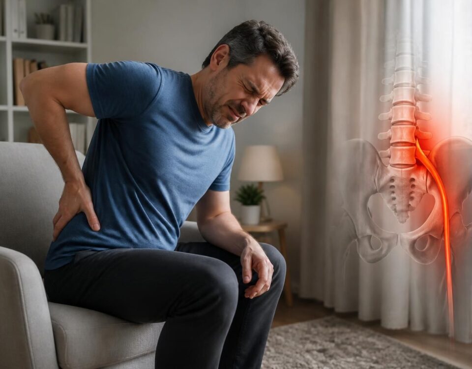
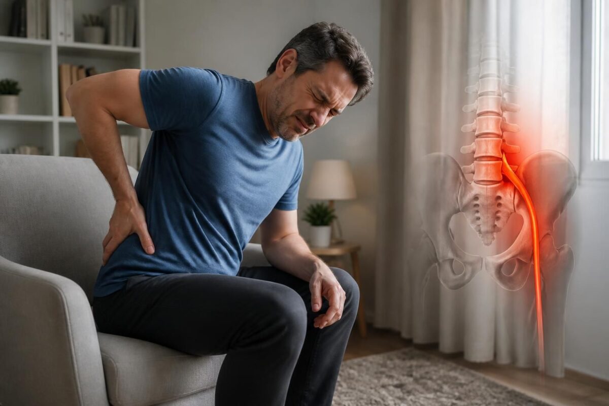
The typical pain associated with sciatica often starts in the lower back and radiates through the buttock, the back of the thigh, the calf, or the foot. The pathway of the pain and its radicular nature (electric, burning, with tingling) point toward an affected nerve root rather than simple muscle-ligament pain. The doctor also explores neurological signs: loss of sensation, weakness in lifting the foot, pain aggravated by coughing, or movements that increase lumbar pressure.
Certain elements require prompt management: marked loss of strength, urinary disturbances or bowel control issues, anesthesia in the genital area, rapidly progressing pain, fever, or compromised general syndrome. These symptoms may signal a risk for the cauda equina and require urgent medical evaluation. The clinical examination, including nerve tension tests and assessment of reflexes and strength, remains the cornerstone of diagnosis and helps avoid hasty diagnoses based solely on an image.
Imaging is not systematic or definitive at the onset of symptoms. MRI becomes useful when pain persists despite appropriate management, when a neurological deficit evolves, or when a specialized intervention is considered. It can specify the exact location of the compression, but it does not replace clinical reasoning: an image may show a herniation, but this is not necessarily the cause of the disorder or its functional extent.
Management is progressive and multidisciplinary: rehabilitation, adapted physical activity, strengthening of core muscles, improvement of postures, and pain management through non-surgical options. Injections may be considered if inflammation around the nerve root persists, but surgery remains a last resort after treatment has failed and in cases of significant neurological deficit.
In practice, the role of the physician is to treat the system, not an isolated image. The gradual return to movement, controlled breathing, improvement of dynamic posture, and reduction of pain-related fear are the most effective levers. Understanding that MRI cannot decide alone helps avoid the trap of quick decisions and opt for an appropriate, patient-centered solution based on real outcomes rather than an isolated picture.
This conclusion highlights that MRI cannot solely determine a diagnosis of disc herniation or sciatica. Effective diagnosis relies on the consistency between symptoms and clinical examination, and imaging primarily serves to locate potential compression to guide treatment, not to decide alone. A progressive approach, including movement, rehabilitation, and appropriate therapeutic options, is generally favored to promote healing and limit unnecessary interventions.
MRI can help visualize structures and locate potential compression, but it does not determine the presence of radicular-origin pain or the severity of a disc-radical conflict on its own. A disc herniation may be visible in individuals without pain, and conversely, a modest image may be associated with significant pain. The diagnosis remains predominantly clinical and contextualized.
The diagnosis primarily relies on clinical evaluation. The medical history and physical examination aim to connect the pain pathway, neurological signs, and tension tests to one or more nerve roots. This approach avoids excessive mechanical interpretations and allows for targeting appropriate treatment rather than relying solely on an image.
The pain pathway and specific signals guide the identification of potentially compressed roots. Pain radiating down the buttock, back of the thigh, calf, or foot suggests sciatica, while more anterior pain may point towards femoral nerve pain. Electric shock sensations, tingling, or pain aggravated by certain movements or coughing strengthen the hypothesis of a disc-radical conflict.
Neurological signs play a key role. Numbness, loss of sensitivity in a specific region, muscle weakness, or difficulty lifting the foot indicate nerve damage and guide the therapeutic strategy. The physician does not rely solely on the location of the pain, but on the extent of the nerve territory it serves.
When MRI is useful and when it can be unnecessary. MRI is particularly useful in cases of persistent pain despite coherent management, neurological deficit, or before a targeted intervention. However, in the majority of sciatica cases that are evolving favorably, it has little value in accelerating healing and may even perpetuate anxiety or limit movement if misinterpreted.
Treatment is progressive and personalized. Typical management combines analgesics and anti-inflammatories when appropriate, activity advice, and rehabilitation focused on movement and postures. Injections may be considered in cases of radicular pain resistant to initial treatment. Surgery remains a last resort option, considered after several weeks of progression with persistent functional impact or in cases of neurological complication.
Avoid pitfalls related to imaging. An image alone is not sufficient to explain pain: it may sometimes indicate a non-painful anomaly or, conversely, underestimate a functional nerve irritation. The nocebo effect can arise when imaging terms are interpreted as definitive verdicts. A system-centered approach, focusing on movement and understanding of pain, promotes recovery.
Additional resources to deepen practical aspects and concepts related to sciatica and disc herniation, including links on chemical irritation of the nerve root, muscle strength, and imaging indications, are available here:
Sciatica Paris,
Chemical irritation of the nerve root,
Muscle strength and why to test it,
Diagnosis and imaging examination,
Stronger pain in the leg,
MRI alone is not enough,
Lumbar osteoarthritis and combined causes of sciatica,
Diagnosis and examination,
Pain radiating lower after a movement,
Warning signs.
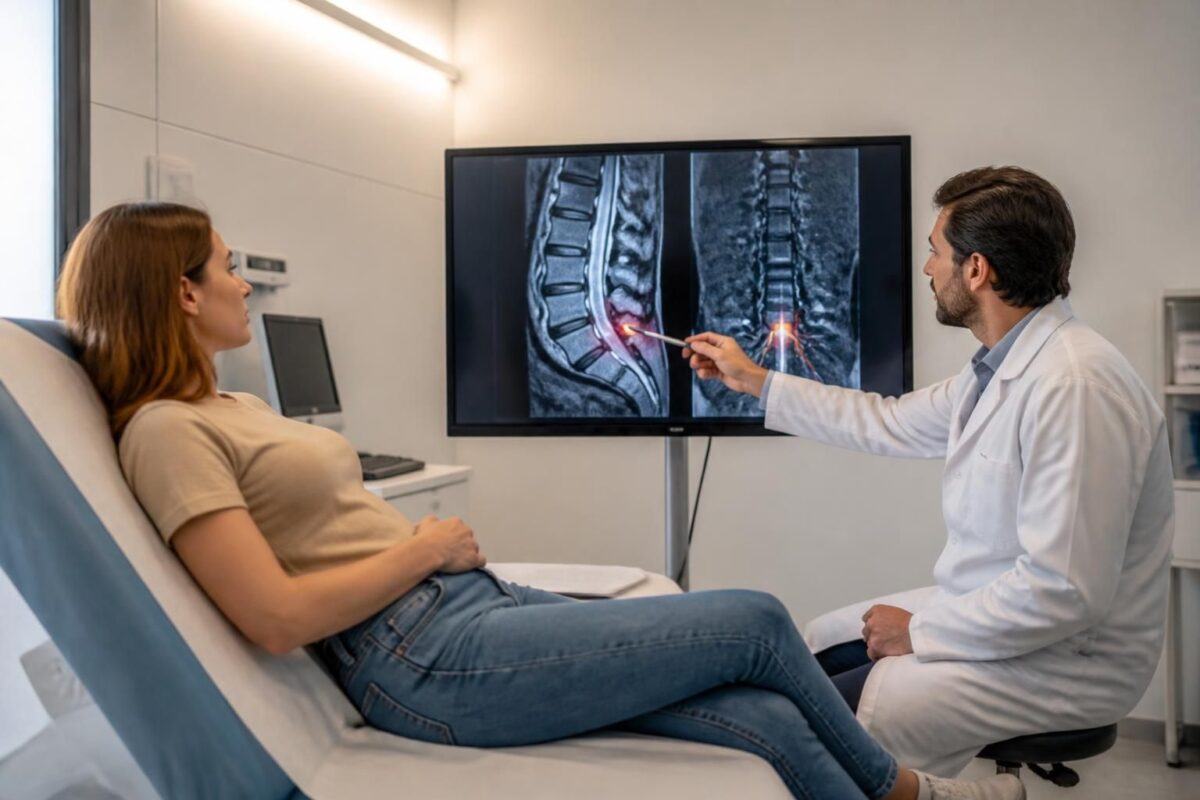
Herniated disc and sciatica: what the MRI can show
IN BRIEF In brief, the MRI is the key tool for evaluating a herniated disc and sciatica. It specifies the location, type (protrusion, extrusion) and degree of compression of the nerve roots, guiding choices for non-surgical or surgical treatment. Its…
Herniated disc and sciatica: why an accurate diagnosis matters
IN SHORT In short, disc herniation and sciatica cause lower back pain and radicular pain. A precise diagnosis identifies the source, localizes the impairment, and guides the most appropriate treatment, avoiding unsuitable treatments. The diagnosis combines a rigorous clinical examination…
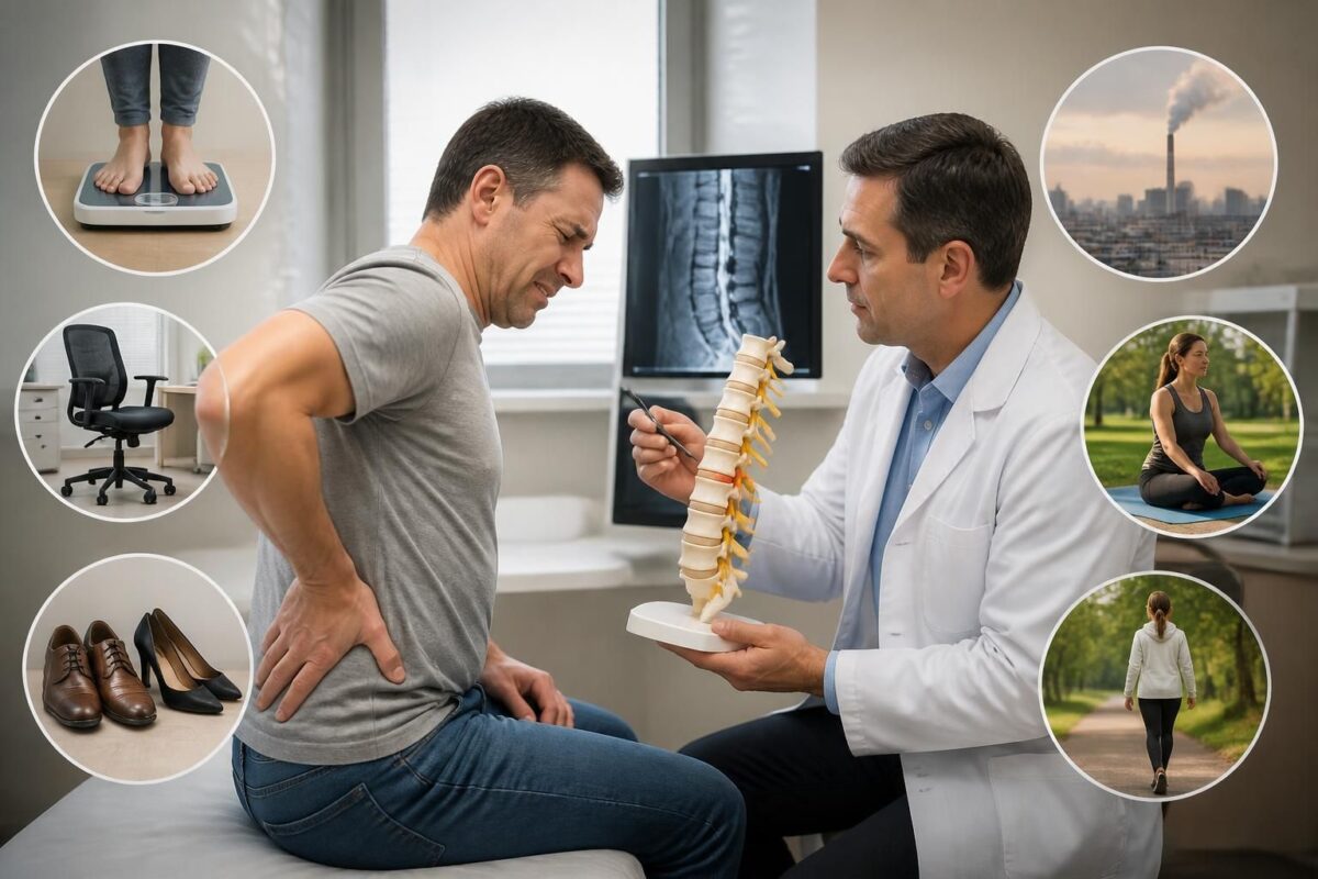
Herniated disc and sciatica: factors that influence progression
IN BRIEF In brief, disk herniation and sciatica are two distinct but often associated mechanisms that depend on a set of mechanical and inflammatory factors. The main advantage of understanding these factors is to guide an adapted and progressive rehabilitation,…
Herniated disc and sciatica: when the pain becomes persistent
IN BRIEF In short, disc herniation and sciatica refer to lower back pain radiating into the leg due to nerve compression. Follow-ups link pain pathway, neurological examination, and relevant imaging to guide management, usually without surgery. Benefits: self-management, maintenance of…

Herniated disc and sciatica: when the cause is mechanical
IN SHORT In short, Herniated disc and sciatica refer to radicular pain due to a mechanical cause when the nucleus of the disc protrudes and irritates a nerve root. The benefits of management include quick pain relief, maintenance of mobility,…
Herniated disc and cauda equina syndrome: warning signs
IN BRIEF In brief, Herniated disc and cauda equina syndrome describe severe compression of the lumbar roots with warning signs requiring urgent management. The warning signs help prevent major sequelae and facilitate rapid referral to emergency services. The benefits include…
Herniated disc and muscle strength: why to test it
IN BRIEF The topic is the herniated disc and muscle strength. Testing muscle strength in this context helps assess the ability of the back and trunk muscles to support the spine and identify weaknesses that may exacerbate pain or limit…

Herniated disc and diminished reflexes: possible neurological sign
IN BRIEF In short, a herniated disc is a partial rupture of the disc ring that can compress a nerve root and cause a neurological sign such as decreased reflexes. This condition presents as pain, often in the lower back…

Dr Sylvain Desforges, B.Sc., D.O., N.D., osteopath, is the founding president of TAGMED and of the ACMA association. He dedicates his practice to chronic spinal pain, low back pain and symptoms compatible with sciatica, particularly when pain radiates toward the buttock, leg, calf, foot or toes.
Through SOS Sciatique, he publishes educational content on sciatica symptoms, possible causes such as disc herniation, disc narrowing, foraminal stenosis or irritation of a lumbar nerve root, as well as non-surgical approaches that may be considered according to the evaluation. At Clinique TAGMED, care may include, depending on the case, motorized neurovertebral decompression, specific osteopathic care or precision percussion.
For an evaluation related to sciatica treatment in Montreal / Mount Royal or sciatica treatment in Terrebonne, please consult the services available at Clinique TAGMED.
Medical disclaimer
The information published on SOS Sciatique is provided for educational purposes only and does not replace a consultation, diagnosis, opinion or treatment provided by a physician or another authorized healthcare professional. The author of this article is neither a medical doctor nor a specialist in a medical specialty recognized by the Collège des médecins du Québec. The osteopathic, manual medicine or functional support approaches described on this site do not constitute medical acts and are not intended to replace medical follow-up.
Consult a physician or emergency service in cases of loss of bladder or bowel control, anesthesia in the saddle area, significant or progressive weakness in the leg or foot, fever, recent trauma, severe pain or rapid worsening of symptoms. For more details, please read our Legal Notice.