


Sciatica and pain when leaning forward
21/09/2026
Sciatica and descending stairs: calf or foot pain
21/09/2026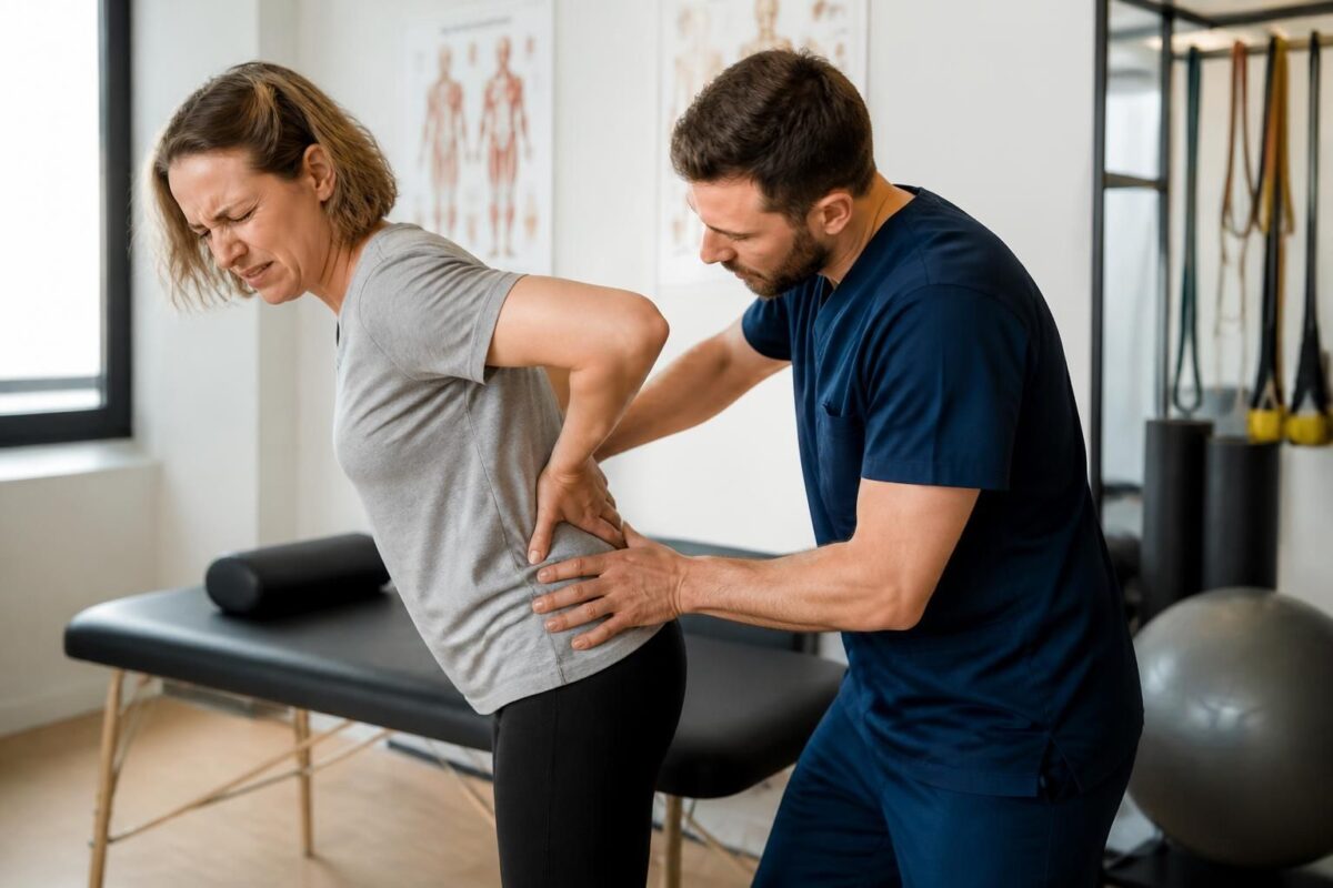
IN SHORT |
| In short, Herniated disc and sciatica refer to radicular pain due to a mechanical cause when the nucleus of the disc protrudes and irritates a nerve root. The benefits of management include quick pain relief, maintenance of mobility, and a targeted rehabilitation that prevents recurrences. The approach is based on a precise clinical diagnosis and a multidisciplinary pathway: examination, imaging if necessary, and conservative approaches (exercises, physical therapy) before any surgery. Available in Montreal, this service aims to quickly return to daily activities while avoiding prolonged immobilization. |
Herniated disc and sciatica: when the cause is mechanical, the pain results from compression of a nerve root by a disc protrusion, often accompanied by local inflammation.
- The sciatic nerve, the longest in the body, can be irritated along the path of the roots, with typical signs according to L5 or S1.
- Path of the roots: L5 radiates to the big toe and the outer side of the thigh; S1 travels down the back of the leg to the heel and the little toe.
- Diagnosis: clinical examination supplemented by an MRI that confirms the location and degree of disc-root conflict.
- Management: conservative approach with controlled movement, physiotherapy, and standard treatments; injections possible if necessary; surgery rare according to neurological deficits.
- Prognosis: over 70–80% improvement in 6–12 weeks; many hernias resolve spontaneously over time.
A progressive and personalized approach is recommended to quickly regain mobility and prevent recurrences.
Quick summary: This article, written by a health expert, explains when herniated disc and sciatica relate to a mechanical cause. It describes the path of the sciatic nerve, typical signs, diagnostic criteria, and non-surgical management options. It highlights situations that require intervention and expected outcomes, drawing on the experience of structures like the TAGMED Clinic and specialized resources.
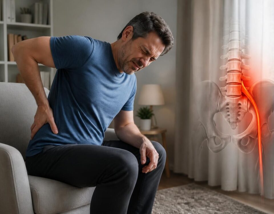
Pain related to a herniated disc is often explained by a mechanical and inflammatory mechanism. The intervertebral disc can deform and compress a nerve root, causing a burning pain or electrical shocks that radiate into the buttock, leg, calf, or foot. To understand the exact path and target the origin of the conflict, one must follow the path of the sciatic nerve, the longest in the body.
The path of the nerve and the pain mechanism
The sciatic nerve originates in the lumbar region and extends to the foot. When a disc deforms and its protrusion irritates the nerve root, the pain follows a precise path according to the affected root. Irritation tends to manifest as burning sensations, electrical shocks, or tingling that extend along the leg. For more in-depth information, you can consult the following resources: PostureA – Herniated Disc and SOS Sciatica – Pain without back pain.
Differentiating L5 and S1
The precise location of pain helps identify the affected root. Lumbar irritation associated with pain that follows the outer path of the thigh, along the ankle, and reaches the big toe typically corresponds to an L5 involvement. Conversely, pain that travels to the back of the leg to the heel and little toe indicates an S1 involvement. Following the path of the nerve to the foot allows for pinpointing the disc-root origin and guiding evaluation and management.
Diagnosis and warning signs
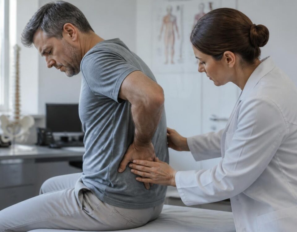
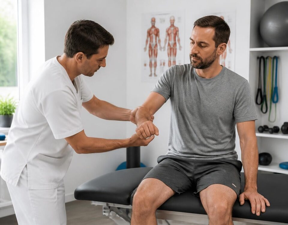
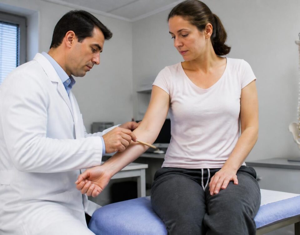
The diagnosis is primarily based on the clinical examination, identifying radicular signs and motor or sensory disturbances. Tests such as the Lasègue and Spurling can help confirm clinical suspicion. Imaging is useful to confirm the diagnosis and assess the extent of disc-root conflict and perineural inflammation. In cases where MRI is contraindicated, a CT scan may be considered, although it is less precise for soft tissues. For certain situations, an EMG may be prescribed. Warning signs such as cauda equina syndrome require urgent evaluation and rapid decompression.
For further information, see resources on SOS Sciatica – Cauda Equina and Foraminal Herniated Disc.
Non-surgical management and options at TAGMED Clinic
In the majority of cases, a non-surgical approach is effective. Treatment is based on multidisciplinary evaluation and an adapted progression, aiming to relieve pain and restore function while avoiding chronicity. At TAGMED Clinic, options may include interventions such as neurovertebral decompression and targeted osteopathy, in addition to a rehabilitation program and inflammation management. Radiologically guided injections may be considered for rapid relief when pain persists despite initial treatment. To access TAGMED services, visit TAGMED Clinic.
Neurovertebral decompression and other non-surgical approaches can be discussed after a comprehensive evaluation. For general information on mechanisms and associated approaches, you can also visit specialized resources such as SOS Herniated Disc and SOS Herniated Disc.
Additional information on the risks of inappropriate use of generic exercises is available at SOS Sciatica – Generic Exercises.
For orientation towards available resources and protocols, you can also consult the information pages on pain that radiates after movement and the pages related to neurovertebral decompression.
When to consider surgery
Surgery is considered in specific cases. It may become necessary when pain persists despite well-managed treatment over several weeks, or in the presence of progressive neurological deficit or cauda equina syndrome. Modern techniques include microsurgical approaches and minimally invasive options tailored to each location. Rigorous post-operative follow-up is essential to consolidate healing and prevent residual pain.
Prognosis and prevention
The prognosis is generally favorable. Many disc herniations can regress spontaneously, partly due to inflammatory resorption mechanisms. Over 70% of patients observe significant improvement within 6 to 12 weeks. In cases of surgery, the success rate for pain relief is high, but long-term monitoring remains necessary to prevent recurrences or chronicization.
Prevention relies on regular physical activity, good ergonomics at work and home, a healthy weight, and adopting proper postures when lifting loads. Tobacco use and poor sleep can promote disc degeneration and spinal pain.
For additional reading on management and clinical distinctions, you can consult the following resources: Tremplin Santé – Clinical Distinctions, Spine Surgery – L5-L4, and ScienceDirect – scientific review.
Additional resources: Disc herniation and pain, Radiating pain after movement, Foramina and root space.
For more information on specialized approaches and clinical resources, also visit the site TAGMED Clinic.
Medical disclaimer: The information and advice provided on this site do not replace the opinion, diagnosis, or treatment of a health professional. Please note that Dr. Sylvain Desforges, osteopath, is neither a medical doctor nor a physician, and is not a specialist in a medical specialty as defined by the Collège des médecins du Québec. Manual medicine, functional medicine, and sports medicine as described on this site exclude any treatment or medical diagnosis performed by a doctor or medical specialist. Always consult your physician for any medical questions. For more details, please read our complete Legal Notice.
- Mecahnical origin: disc herniation compressing a lumbar root
- Nerve pathway: pain following L5 or S1
- Signs: radiculalgia, burning, electric shock sensation
- Pathway: pain towards the foot following the pathway
- Diagnosis: MRI to visualize disc-root conflict
- Management: preference for non-surgical
- Treatment: light movements, physiotherapy, core stabilization
- Inflammation: Medications and, if necessary, injections
- Emergency: signs of cauda equina or progressive weakness
- Prevention: posture, physical activity, ergonomics

This article provides practical recommendations when a disc herniation causes a sciatica of mechanical origin. It describes the mechanism, the typical paths of the sciatic nerve, non-surgical management approaches, and rehabilitation gestures to prioritize to regain mobility, while recognizing warning signals that require prompt evaluation.
Understanding the mechanism and pathway of the nerve
The sciatic nerve is the longest in the body and can be irritated when the nucleus pulposus of the intervertebral disc migrates through the fibrous ring. This mechanical compression is accompanied by a local inflammatory reaction, generating intense pain that radiates along the paths of the L5 or S1 roots. In practice, an L5 injury often manifests on the outer thigh and then the big toe, while an S1 injury follows the back of the leg to the heel and the little toe. Understanding this pathway helps target the discal origin and guide rehabilitation.
Signs of injury and clinical markers
Typical mechanical origin symptoms include low back pain associated with radiation to the leg, tingling or burning sensations, and sometimes muscle weakness. Pain is usually triggered or aggravated by prolonged sitting, bending efforts, and episodes of coughing or sneezing. MRI remains the key examination to confirm a disc protrusion and assess the exact location. In practice, the presence of marked neurological deficits or muscle weakness requires prompt management and may guide an adapted approach between physiotherapy and, in certain cases, a more targeted intervention.
Non-surgical management and posture
For most mechanical cases, non-surgical treatment is effective. Strict rest is not recommended; instead, prioritize suitable physical activity that promotes vascularization and recovery. Anti-inflammatories or pain relievers may be used for short periods as assessed by the professional, with an approach tailored to the patient’s profile. The goal is to reduce inflammation, preserve mobility, and avoid repeated crises through a gradual increase in daily and professional activities.
Rehabilitation and Key Exercises
Rehabilitation is central to restoring mobility and strengthening the deep trunk muscles. A personalized program may combine core strengthening, lumbar strengthening, proprioception exercises, and targeted stretches of the hamstrings and pelvic-peritoneal muscles. The McKenzie method and neuro-myelogenic mobilization techniques may be offered to gradually restore lumbar mobility and decrease radicular irritation. Gradually, the goal is to increase endurance, improve posture, and prevent recurrences while remaining attentive to the patient’s tolerance and symptom evolution.
Examples of Exercises and Progression
Start with gentle and appropriate exercises, then increase the difficulty as improvements occur. Include diaphragmatic breathing exercises, trunk strengthening exercises, and progressive trunk flexion and extension movements. Integrate pelvic stability exercises and “plank” series on the knees or forearms, ensuring to maintain good technique and avoid acute pain. Professional supervision is recommended to tailor the plan to your level and pain.
When to Consider Intervention and Monitoring
Surgery is considered in rare cases when pain persists despite optimized treatment over several weeks, or in the presence of a progressive neurological deficit, or cauda equina syndrome. After any intervention, monitoring and rehabilitation remain essential to consolidate the lumbar area and reduce the risk of recurrence.
Prevention and Continuity of Care
To prevent recurrences, maintain regular physical activity, adopt ergonomics suitable for work and home, and prioritize good postural hygiene when lifting loads. Avoid aggravating factors like tobacco and ensure restorative sleep. Regular follow-up with a professional allows for adjusting the program and sustainably supporting functional recovery.
| Criterion | Concise Text |
|---|---|
| Definition | Disc herniation refers to the extrusion of the nucleus of the disc that compresses a nerve root and triggers radicular pain when the cause is mechanical. |
| Mechanism | Pain associated with a mechanical phenomenon and local inflammation around the root. |
| Location | Lower back pain with possible radiation to the lower leg depending on the affected root (L5–S1 most common). |
| Pain Pathway | Follows the pathway of the sciatic nerve with variations depending on L5 or S1. |
| Imaging | MRI is the reference examination for visualizing the herniation and the disc-root conflict. |
| Initial Management | Light physical activity, rehabilitation, and pain relief or short-term NSAIDs. |
| Specific Approaches | Targeted infiltrations or techniques such as ozonotherapy may be considered in case of persistence. |
| Emergencies | Surgical emergency in case of cauda equina syndrome or progressive motor loss. |
| Prognosis | Most evolve favorably; resorption is possible and rehabilitation accelerates the return to activity. |
Testimonials: Disc Herniation and Sciatica – When the Cause is Mechanical
My name is Marie, 41 years old, and my daily life changed after a simple lift at work. What seemed like lower back pain quickly turned into a disc herniation and a sciatica of mechanical origin. Thanks to a rehabilitation program focused on movement and targeted sessions, the pain has decreased and my mobility has returned.
I’m Nicolas, 48 years old, and my daily life was blocked by lower back pain radiating along my thigh to my big toe. The diagnosis is a disc herniation with sciatica of mechanical origin. With appropriate exercises and corrected posture, I regained my ability to work and move normally.
I’m Sophie, 34 years old, and my issue arose from poor posture at the office. The sciatica appeared with radicular pain and a disc herniation due to a mechanical mechanism. A progressive rehabilitation plan allowed me to resume my sports activities without pain.
My name is Pierre, 57 years old, and my life was turned upside down by lower back pain radiating to my buttock and leg. The verdict: disc herniation due to a mechanical mechanism. After a structured course and strengthening exercises, I regained mobility and can now resume my daily tasks without limitation.
I’m Julie, 29 years old, an athlete and avid walker. My sciatica was linked to a disc herniation with a mechanical mechanism, causing electric shocks when sitting. Thanks to a precise assessment and a gradual rehabilitation plan, I am progressively regaining my performance, and my daily routes are now pain-free.
Dr. Sylvain Desforges, osteopath and expert in manual medicine, adopts a pragmatic and deeply human approach to spinal pain. His practice is based on a strong conviction: most cases of sciatica and herniated discs stem from a mechanical mechanism triggered by years of postural stress and microtrauma. By prioritizing a precise understanding of anatomy and the path of the longest nerve in the body, he guides his patients towards care pathways that combine effectiveness and safety, without unnecessarily resorting to invasive interventions.
At the heart of this issue, the anatomy of the sciatic nerve proves to be decisive. At its widest point, near the pelvis, the impressive diameter of the nerve makes it a vulnerable target for compression and irritation. A herniated disc can project the nucleus pulposus toward the annulus fibrosus, causing root irritation that manifests as burning sensations and electric shocks, especially during prolonged sitting or after sudden exertion. Understanding this path allows for quick differentiation between inflammatory aggravation and potential surgical emergencies.
The distinction between the lesions of roots L5 and S1 is not merely theoretical: it influences treatment and exercises. Pain that follows the external path of the thigh, curves around the malleolus to target the big toe often signals an L5 lesion, while a path that ascends the back of the leg to the heel and the little toe points to S1. Following this pathway to the foot enables precise identification of the disc-radicular origin and avoids misalignment in treatment.
Contrary to the old adage of strict rest, pain related to a disc compression and inflammation is now better understood through adapted movement. The disc releases chemical mediators that irritate tissues and nerves; thus, maintaining continuity of light physical activities, under supervision, promotes blood circulation and speeds up recovery. In most cases, the natural resorption process can start within weeks and extend over several months, with a key role for rehabilitation and strengthening of the core muscles.
The care pathway he favors relies on a multidisciplinary and progressive approach. After a thorough clinical evaluation and, if necessary, imaging assessment, Dr. Desforges prioritizes non-surgical solutions focused on maintaining movement, physiotherapy, and targeted manual techniques. When necessary, complementary modalities such as motorized neurovertebral decompression, specific osteopathy, and precision tools may be integrated to reduce pain and restore mobility while respecting the structure of the spine.
The statistics remain reassuring: a majority of herniated discs resolve spontaneously, and over 70% of patients see significant improvement within 6 to 12 weeks thanks to appropriate care. Dr. Desforges stresses the importance of a personalized plan that combines therapeutic education, posture, proprioception, and core-strengthening exercises, in order to minimize recurrences and optimize the return to an active and pain-free life.
With a career dedicated to innovation in pain care, he strives to integrate advanced technologies and evidence-based approaches to optimize the quality of life for his patients. His goal is clear: to help each person regain enduring mobility, prioritizing safety and effectiveness. To deepen his approach and discover the services offered, patients turn to TAGMED clinics, where expertise and personalized support make a difference.
This text summarizes the interactions between herniated discs and sciatica when the cause is mechanical, emphasizing the path of the sciatic nerve, key signs, and treatment options focused on movement and rehabilitation. It explains how to differentiate lesions of roots L5 and S1, describes the inflammatory mechanisms accompanying pain, and proposes current criteria for assessing emergencies and guiding the care pathway in 2025.
In the mechanical framework, pain arises from a mix of compression and inflammation around the sciatic nerve. A herniated disc can irritate the nerve root and trigger burning sensations or electric shocks that follow a specific path depending on the affected root. Understanding this pathway helps to target the diagnosis and guide treatments towards an effective and lasting resolution.
Movement remains the guiding principle of relief. Contrary to the notion of absolute rest, an adapted activity promotes tissue vascularization and accelerates recovery. Rehabilitation programs focused on strengthening deep muscles, core stability, and neuro-meningeal mobilization restore lumbar mobility and prevent recurrences, with special attention given to the specific indications related to sciatica and low back pain.
Urgency scenarios remain infrequent but crucial to recognize. Persistent ultracratique pain, progressive loss of strength, or signs such as saddle anesthesia and sphincter disorders require rapid assessment, even surgical decompression in the most severe cases (cauda equina syndrome). Surgical intervention is then decided based on clear criteria when conservative management does not provide relief or when neurological deficits rapidly worsen.
After any potential intervention, rehabilitation remains central. It aims to solidify lumbar support, correct postures, and restore optimal daily activities while reducing the risk of recurrence. The goal is a sustainable active return to activities, with appropriate monitoring to prevent residual pain or postoperative complications.
The prevention of recurrences involves a combination of lifestyle and posture hygiene. Regular physical activity, weight management, ergonomics tailored to work and home, smoking cessation, and good sleep quality play a key role in reducing episodes of lumbar and radicular pain. At the same time, therapeutic education encourages simple daily gestures to keep tissues supple and strong.
To deepen your understanding of the mechanisms and nerve pathways associated with herniated discs and sciatica, consult the following resources: Herniated disc and foot weakness, Herniated disc and nerve pathway, Chemical irritation of the nerve root, Inflammation of the nerve root, Acute sciatica – causes and frequencies, Understanding herniated discs and sciatica, IRM warning signs and herniated disc, Recurrence of sciatica – possible factors, Scientific study on herniated disc.
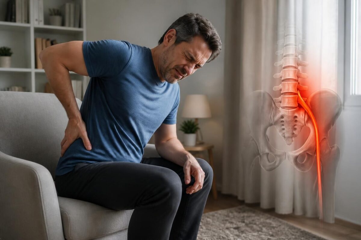
Herniated disc and sciatica: when the pain becomes persistent
IN BRIEF In short, disc herniation and sciatica refer to lower back pain radiating into the leg due to nerve compression. Follow-ups link pain pathway, neurological examination, and relevant imaging to guide management, usually without surgery. Benefits: self-management, maintenance of…
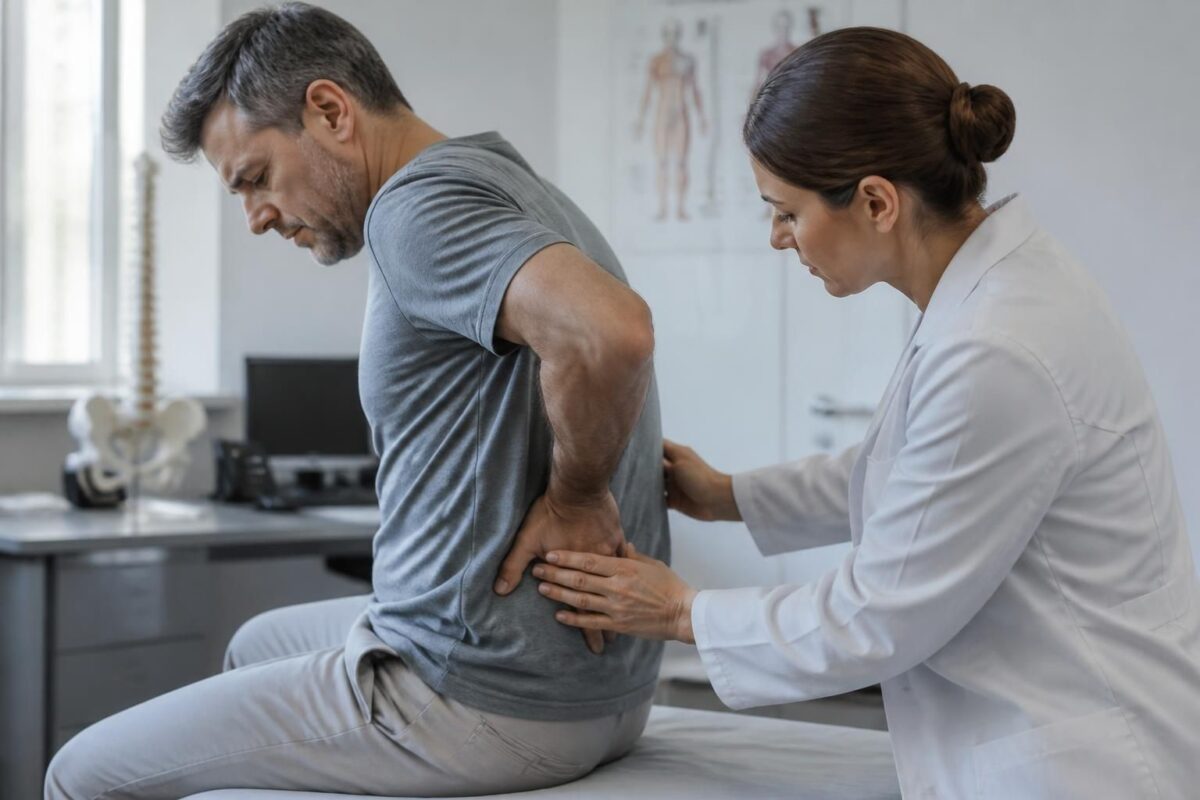
Herniated disc and cauda equina syndrome: warning signs
IN BRIEF In brief, Herniated disc and cauda equina syndrome describe severe compression of the lumbar roots with warning signs requiring urgent management. The warning signs help prevent major sequelae and facilitate rapid referral to emergency services. The benefits include…
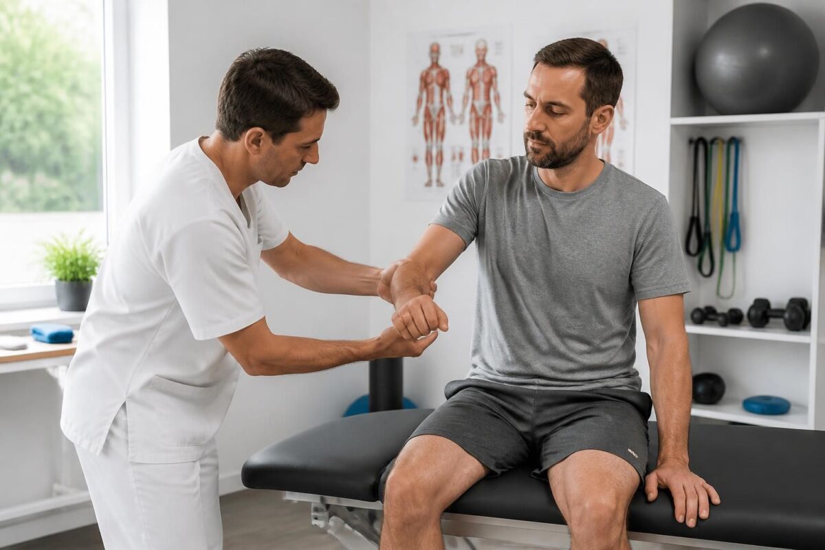
Herniated disc and muscle strength: why to test it
IN BRIEF The topic is the herniated disc and muscle strength. Testing muscle strength in this context helps assess the ability of the back and trunk muscles to support the spine and identify weaknesses that may exacerbate pain or limit…

Herniated disc and diminished reflexes: possible neurological sign
IN BRIEF In short, a herniated disc is a partial rupture of the disc ring that can compress a nerve root and cause a neurological sign such as decreased reflexes. This condition presents as pain, often in the lower back…
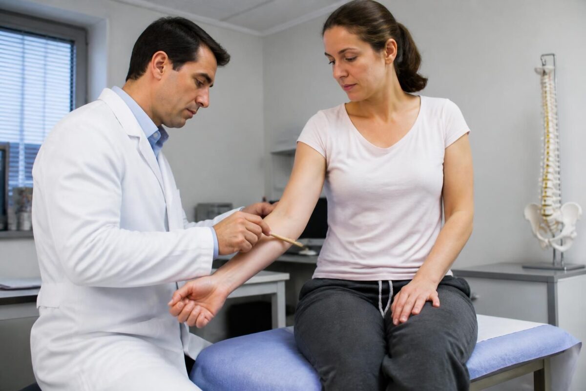
Herniated disc and loss of sensation: why evaluate
IN BRIEF A herniated disc is a protrusion of the gelatinous nucleus of an intervertebral disc that can compress a nerve root, causing radicular pain and a loss of sensation in an area of the body. Quickly assessing this condition…

Herniated disc and foot pain: L5 or S1?
IN BRIEF In brief, Disc herniation and foot pain related to the L5-S1 level explain how a hernia can irradiate towards the foot and how to determine if the pain originates from L5 or S1. This approach aims for a…
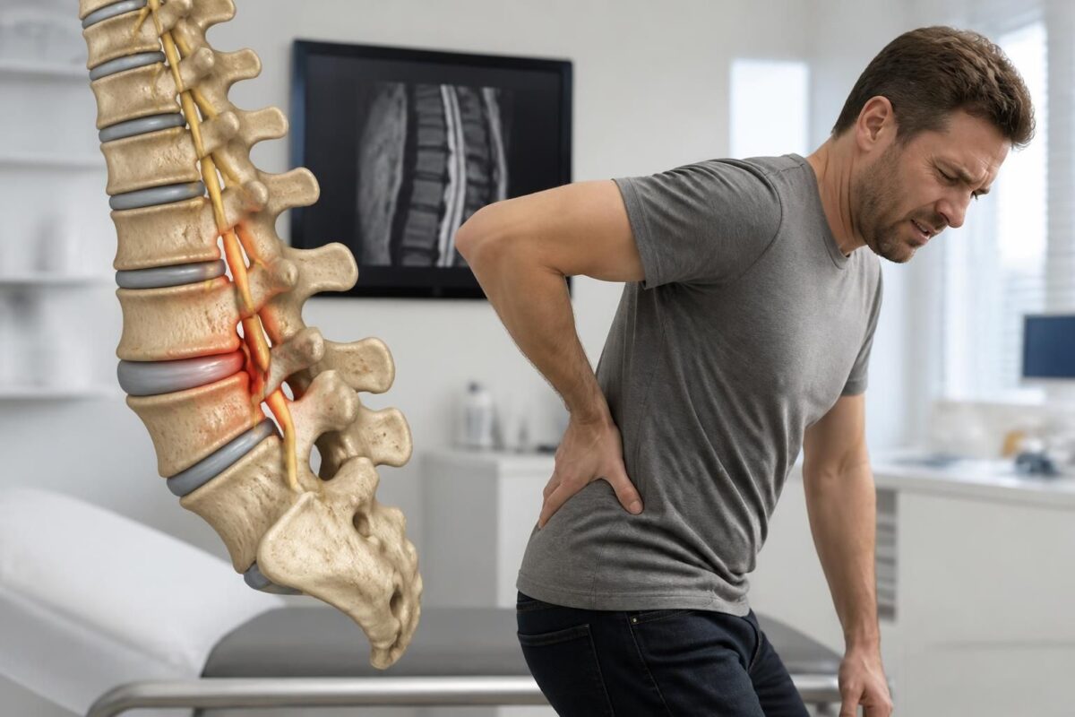
Herniated disc and spondylolisthesis: possible nerve irritation
IN BRIEF The lumbar disc herniation and spondylolisthesis refer to a possible compression of the nerve roots leading to sciatic pain and sometimes paresthesia or muscle weakness. This nerve irritation can mostly be managed through conservative measures and medication, providing…

Herniated disc and lumbar osteoarthritis: combined causes of sciatica
IN BRIEF The subject is the association between herniated disc and lumbar osteoarthritis as combined causes of sciatica and femoral neuralgia. This interaction explains the low back pain and nerve irradiations. Benefits and applications: allows differentiation of pain related to…

Dr Sylvain Desforges, B.Sc., D.O., N.D., osteopath, is the founding president of TAGMED and of the ACMA association. He dedicates his practice to chronic spinal pain, low back pain and symptoms compatible with sciatica, particularly when pain radiates toward the buttock, leg, calf, foot or toes.
Through SOS Sciatique, he publishes educational content on sciatica symptoms, possible causes such as disc herniation, disc narrowing, foraminal stenosis or irritation of a lumbar nerve root, as well as non-surgical approaches that may be considered according to the evaluation. At Clinique TAGMED, care may include, depending on the case, motorized neurovertebral decompression, specific osteopathic care or precision percussion.
For an evaluation related to sciatica treatment in Montreal / Mount Royal or sciatica treatment in Terrebonne, please consult the services available at Clinique TAGMED.
Medical disclaimer
The information published on SOS Sciatique is provided for educational purposes only and does not replace a consultation, diagnosis, opinion or treatment provided by a physician or another authorized healthcare professional. The author of this article is neither a medical doctor nor a specialist in a medical specialty recognized by the Collège des médecins du Québec. The osteopathic, manual medicine or functional support approaches described on this site do not constitute medical acts and are not intended to replace medical follow-up.
Consult a physician or emergency service in cases of loss of bladder or bowel control, anesthesia in the saddle area, significant or progressive weakness in the leg or foot, fever, recent trauma, severe pain or rapid worsening of symptoms. For more details, please read our Legal Notice.